
- Most SCCs are curable with surgery alone — but 2–5% metastasize, and high-risk subsets identified by BWH staging carry nodal metastasis rates of 21–67%, making accurate risk stratification essential
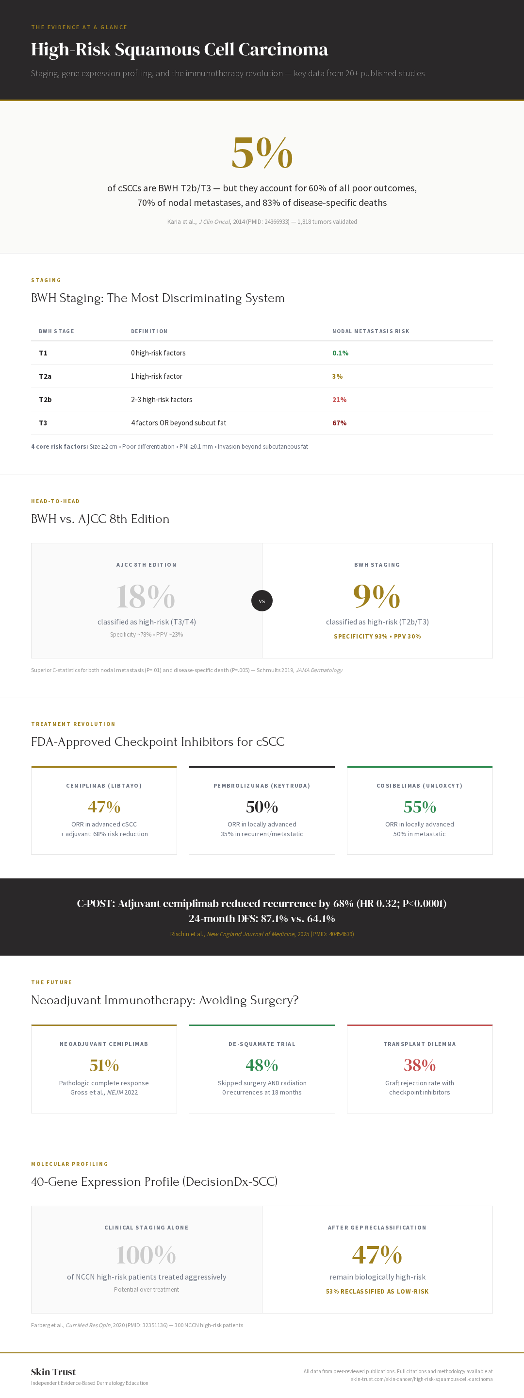
- Three staging systems exist and they disagree — BWH is best at identifying dangerous tumors (5% of cases capture 60% of bad outcomes), NCCN is best for clinical decisions, and AJCC is required for registries and insurance
- Gene expression profiling can reclassify over half of "high-risk" patients as biologically low-risk — but validation concerns remain, and no randomized trial has proven GEP-guided management improves outcomes
- Three FDA-approved checkpoint inhibitors now exist for cSCC — cemiplimab's adjuvant C-POST trial showed a 68% reduction in recurrence, and neoadjuvant immunotherapy achieves pathologic complete response in approximately 50% of patients
- Immunosuppressed patients face dramatically higher risk — organ transplant recipients develop SCC at 20- to 65-fold higher rates, with more aggressive disease, yet most staging systems don't formally account for immunosuppression
- The field is evolving rapidly — neoadjuvant immunotherapy may allow some patients to avoid surgery entirely, but early-phase data requires confirmation in larger trials
This resource is updated monthly as new evidence, staging refinements, and treatment data emerge. High-risk cutaneous squamous cell carcinoma is one of the most rapidly evolving areas in dermatologic oncology — and the goal of this page is to keep patients and providers current. Last updated: March 2026.
Evidence Snapshot
| Key Finding | Data |
|---|---|
| Metastasis Rate (Overall cSCC) | 2–5% of all cutaneous SCCs (Karia et al., 2014); 1.9% in the largest modern registry (Netherlands, 11,137 patients) |
| BWH T2b Nodal Metastasis | 21% (vs. 0.1% for T1) — only 5% of tumors capture 60% of bad outcomes (Karia et al., 2014) |
| GEP Reclassification | 53% of NCCN "high-risk" patients reclassified as biologically low-risk (Farberg et al., 2020) |
| Adjuvant Cemiplimab (C-POST) | 68% reduction in recurrence; 24-month DFS 87.1% vs. 64.1% placebo (Rischin et al., 2025) |
| Neoadjuvant pCR Rate | ~50% pathologic complete response with preoperative cemiplimab (Gross et al., 2022) |
| Transplant SCC Risk | 20- to 65-fold increased incidence in organ transplant recipients |
What Makes a Squamous Cell Carcinoma "High-Risk"?
Most squamous cell carcinomas are straightforward to treat. A skilled Mohs surgeon removes the tumor, confirms clear margins under the microscope, repairs the wound, and the patient goes home the same day with a cure rate approaching 99%. The problem is the small but consequential minority of SCCs that don't follow the script — the ones that recur, invade nerves, metastasize to lymph nodes, and occasionally kill.
These are high-risk squamous cell carcinomas, and they represent one of the most confusing areas in all of dermatologic oncology. Even among expert Mohs surgeons, there is genuine uncertainty about how to stage these tumors, when to recommend additional testing, whether sentinel lymph node biopsy adds value, and how aggressively to pursue adjuvant treatment. The staging systems disagree with each other. The gene expression profiling data is promising but debated. And the immunotherapy revolution has introduced treatment options that didn't exist five years ago.
This article exists because this confusion matters. A patient with a high-risk SCC deserves to understand their situation clearly, and a physician managing one deserves a single, current, evidence-based resource that cuts through the noise.
Who Gets High-Risk Squamous Cell Carcinoma?
Cutaneous SCC is the second most common cancer in the United States, with approximately 1.8 million cases annually. Men develop SCC roughly two to three times more often than women, and incidence rises sharply after age 50.
Demographics and epidemiology. Cutaneous SCC disproportionately affects older, fair-skinned men with cumulative UV exposure. The incidence has been rising steadily over the past several decades, driven by an aging population, increased sun exposure, and improved detection. Immunosuppressed patients — particularly organ transplant recipients — face a dramatically elevated risk, developing SCC at an estimated 20- to 65-fold higher rate than the general population (with some older reviews citing figures as high as 250-fold depending on the immunosuppressive regimen and duration), often with more aggressive disease and higher recurrence rates.
The immunosuppression problem. Organ transplant recipients represent a special population in high-risk SCC. These patients develop SCCs earlier, more frequently, with more aggressive histologic features, and with substantially worse outcomes stage-for-stage compared to immunocompetent patients. Critically, neither the AJCC 8th edition nor the BWH staging system formally incorporates immunosuppression status — a gap that the NCCN guidelines attempt to address by listing immunosuppression as an independent high-risk factor. In my experience treating immunosuppressed patients, these tumors demand a fundamentally different level of vigilance. A T1 SCC in a transplant recipient is not the same disease as a T1 SCC in a healthy 70-year-old. A 2024 prospective multicenter study (SCOPE-ITSC) by de Jong et al. found organ transplant recipients with high-stage, ulcerated cSCCs had a 46.2% 2-year cumulative incidence of metastasis — a sobering reminder that risk stratification in this population requires heightened vigilance.
Chronic lymphocytic leukemia (CLL) patients also carry markedly elevated SCC risk and worse outcomes, as do patients on long-term immunosuppressive therapy for autoimmune conditions.
Your immune system is your body's surveillance camera for cancer. It catches and kills abnormal cells before they become a problem. Organ transplant patients take drugs that deliberately turn down that surveillance system so their body doesn't reject the transplanted organ. The tradeoff is real: you keep your kidney, but your skin cancer risk skyrockets — and the cancers that do develop tend to be meaner because there's no immune system keeping them in check. This is why I treat a "low-stage" SCC in a transplant patient with the same seriousness I'd treat a higher-stage tumor in someone with a normal immune system. The staging systems haven't caught up to this reality yet.
— Dr. Hocker
What Are the Risk Factors That Make an SCC High-Risk?
The distinction between a routine SCC and a high-risk SCC is determined by a combination of tumor-specific and patient-specific factors. Understanding these is essential because they drive every downstream decision — from staging to surveillance to treatment.
Tumor-Specific High-Risk Features
Size ≥2 cm. Tumor diameter is one of the most consistently validated risk factors across all staging systems. SCCs measuring 2 cm or larger have significantly higher rates of local recurrence, nodal metastasis, and disease-specific death.
Poor differentiation (Broder's grade 3–4). Histologically, poorly differentiated SCCs show minimal keratinization, marked nuclear pleomorphism, and high mitotic rates. They are harder to diagnose and behave more aggressively. The Broder's grading system classifies tumors on a 1–4 scale, with grades 3 and 4 carrying the highest risk. In practice, pathologists typically dichotomize this as "well-to-moderately differentiated" (low-grade) versus "poorly differentiated" (high-grade).
Perineural invasion (PNI). This is among the strongest predictors of poor outcome in cSCC. PNI occurs in 2.5–14% of all cSCCs, but when present — particularly large-caliber PNI involving nerves ≥0.1 mm in diameter — the prognosis changes substantially. Local recurrence rates with PNI range from 16–45%, nodal metastasis from 10–50%, and disease-specific death from clinical PNI approaches 30%. In my personal experience, perineural invasion is the single feature that most reliably separates routine SCCs from dangerous ones.
Think of nerves as highways running through your tissue. Most skin cancers just sit in one spot — they're a pothole in the road. Perineural invasion means the cancer has figured out how to travel along the highway. That's a fundamentally different problem. A small, well-behaved SCC on your temple is usually a one-and-done Mohs case. Add perineural invasion and now I'm worried about where that highway leads — to the brain, the eye, the jaw. It changes everything: the surgery gets bigger, we consider radiation, and surveillance tightens dramatically.
— Dr. Hocker
Depth of invasion. Tumors invading beyond the subcutaneous fat (Clark level V) or deeper structures carry substantially elevated risk. The AJCC 8th edition uses a 6 mm depth threshold as a staging criterion, while the BWH system defines invasion beyond subcutaneous fat as one of its four core risk factors.
Lymphovascular invasion. Though less common than PNI, lymphovascular invasion is an independent predictor of nodal metastasis and poor outcome.
Desmoplastic histology. Desmoplastic SCC is a recognized high-risk variant with a propensity for perineural invasion and local recurrence.
Anatomic High-Risk Sites
The location of the tumor matters independently of its histologic features. The ear, temple, lip, and anogenital region are consistently identified as high-risk anatomic sites across multiple staging systems and guidelines.
Patient-Specific Risk Factors
Immunosuppression (as discussed above) remains the most powerful patient-level risk factor. Recurrent tumors — SCCs that have already recurred after prior treatment — also carry substantially worse prognosis than primary tumors. Notably, neither immunosuppression nor recurrence status is captured by the AJCC 8th edition staging system, which is a major limitation.
How Are High-Risk Squamous Cell Carcinomas Staged?
This is where the confusion begins — and where it matters most. Multiple staging systems exist for cSCC, and they don't always agree. Each uses different criteria, defines risk differently, and has different strengths and weaknesses. A tumor that is "high-risk" by NCCN criteria might be T1 by AJCC and T2a by BWH. Understanding these systems and how they compare is essential for making informed decisions.
AJCC 8th Edition TNM Staging (2017)
The American Joint Committee on Cancer staging system is the most widely recognized system in oncology, and the 8th edition (current) made important changes for cSCC.
T-Stage Criteria:
| Stage | Criteria |
|---|---|
| T1 | Tumor ≤2 cm in greatest dimension |
| T2 | Tumor >2 cm to ≤4 cm |
| T3 | Tumor >4 cm, OR minor bone erosion, OR perineural invasion, OR deep invasion (>6 mm) |
| T4 | Tumor with gross cortical bone/marrow invasion, skull base invasion, or skull base foramen involvement |
What changed from AJCC 7th edition: The 8th edition introduced a 6 mm depth threshold (replacing the old 2 mm/Clark level IV criteria), which dramatically improved staging accuracy. Under AJCC-7, only 0.7% of tumors were classified as T3/T4, missing most high-risk disease. Under AJCC-8, 17.8% are classified as T3/T4, and these account for 70.4% of all poor outcomes.
Nodal staging (N):
| Stage | Criteria |
|---|---|
| N0 | No regional nodal metastasis |
| N1 | Single ipsilateral node ≤3 cm |
| N2 | Single ipsilateral node >3 cm, or multiple ipsilateral, or bilateral nodes |
| N3 | Any node >6 cm |
Limitations: The AJCC 8th edition does NOT incorporate immunosuppression status, does not account for recurrence history, and its nodal staging concentrates a disproportionate number of patients (>90%) into the N2–N3 categories, limiting its ability to discriminate risk among patients with nodal disease.
Brigham and Women's Hospital (BWH) Staging
The BWH staging system, developed by Chrysalyne Schmults and Pritesh Karia at Brigham and Women's Hospital, has emerged as the most discriminating staging system for cSCC in multiple head-to-head comparisons.
The Four Core Risk Factors:
- Tumor diameter ≥2 cm
- Poor differentiation (Broder's grade 3–4)
- Perineural invasion of nerve ≥0.1 mm diameter
- Invasion beyond subcutaneous fat
BWH T-Stage Classification:
| Stage | Definition | Nodal Metastasis Risk |
|---|---|---|
| T1 | 0 high-risk factors | 0.1% |
| T2a | 1 high-risk factor | 3% |
| T2b | 2–3 high-risk factors | 21% |
| T3 | 4 risk factors OR invasion beyond subcutaneous fat | 67% |
In the landmark Karia 2014 validation study of 1,818 cSCCs, BWH T2b and T3 tumors represented only 5% of all cases but accounted for 60% of all poor outcomes, 70% of nodal metastases, and 83% of disease-specific deaths. This is staging at its most efficient — identifying the dangerous minority without over-classifying the safe majority.
BWH vs. AJCC-8 head-to-head (Schmults 2019, JAMA Dermatology): BWH demonstrated superior C-statistics for both nodal metastasis (P=.01) and disease-specific death (P=.005). BWH classified only 9% of tumors as high-risk (T2b/T3) compared to AJCC-8's 18% (T3/T4), meaning BWH achieves better discrimination with fewer patients flagged — higher specificity (93% vs. ~78%) and higher positive predictive value (30% vs. ~23%) for metastasis or death.
2025 refinement: A 2025 JAMA Dermatology study (Ran et al., 2025) further showed that tumors with 3 risk factors have significantly worse outcomes than those with 2 risk factors despite both being classified as BWH T2b, suggesting potential for further subdivision within the T2b category to improve risk stratification.
NCCN Risk Stratification (Updated 2022)
The National Comprehensive Cancer Network takes a fundamentally different approach: instead of TNM staging, it stratifies tumors into three risk tiers.
Low-Risk cSCC:
- Tumor ≤2 cm, well-differentiated, no PNI, no LVI, immunocompetent, primary (not recurrent)
High-Risk cSCC:
- Any of: >2 cm, poorly differentiated, any PNI, LVI, immunocompromised, recurrent, or high-risk anatomic site (ear, temple, anogenital, eyelid, scalp)
Very-High-Risk cSCC (added 2022):
- Tumor >4 cm, deep invasion beyond subcutaneous fat, large-caliber PNI, T3/T4, in-transit metastases
Validation (JAMA Dermatology 2023): A cohort of 8,727 patients (10,196 primary cSCCs) confirmed that the NCCN very-high-risk category significantly enriches for local recurrence, metastasis, and disease-specific death.
Key advantage over AJCC and BWH: NCCN is the only major system that explicitly incorporates immunosuppression and recurrence history. For immunosuppressed patients, this matters enormously.
N1S3 Staging (Nodal Disease)
For patients who have already developed nodal metastases, the N1S3 system (Forest et al., 2010) provides better risk stratification than AJCC nodal staging. It categorizes patients by number and size of involved nodes and shows more balanced distribution across risk categories than AJCC-8, which concentrates >90% of node-positive patients into stage IV.
Staging Systems Comparison Table
| System | Categories | Incorporates Immunosuppression? | Incorporates Recurrence? | Best Validated For | Key Limitation |
|---|---|---|---|---|---|
| AJCC 8th Ed | T1–T4, N0–N3, M0–M1 | No | No | Wide oncologic use; insurance/registry | Poor nodal discrimination; misses patient factors |
| BWH | T1, T2a, T2b, T3 | No | No | Discriminating high-risk primary tumors | Single-institution derivation |
| NCCN | Low / High / Very-High | Yes | Yes | Clinical decision-making | Less granular than TNM |
| N1S3 | I, II, III (nodal only) | No | No | Node-positive head/neck cSCC | Nodal disease only |
| Broder's | Grade 1–4 | No | No | Histologic differentiation | Subjective; inter-observer variability |
No single staging system captures everything. The BWH system is the most discriminating for predicting which primary tumors will metastasize. NCCN is the most comprehensive for guiding clinical decisions because it incorporates immunosuppression and recurrence. AJCC remains the lingua franca for tumor registries and insurance. A complete assessment should consider all three.
Making Sense of the Staging Confusion
If you've read the sections above and feel confused about staging, you're not alone — and the confusion isn't your fault. The staging situation in high-risk SCC is genuinely messy, even for specialists. Here's why, and what you should take away from it.
Why do three different staging systems exist? Because they were built by different groups, at different times, to answer different questions. Think of it like three different GPS apps giving you three different routes to the same destination. Google Maps (AJCC) is the one everyone has — it's the default, it talks to your insurance company, and it's what the tumor registry uses. But it wasn't designed specifically for skin cancer, so it sometimes gives you a route that doesn't make sense for your particular trip. Waze (BWH) was built by skin cancer specialists who studied which tumors actually kill people, and it's the best at predicting which specific tumors are dangerous — but it only looks at the tumor itself and ignores your personal health factors. Apple Maps (NCCN) takes a broader view — it factors in whether you're immunosuppressed, whether this is a recurrent tumor, and gives you a practical risk tier. But it's less granular than the others.
Why do the systems disagree on the same tumor? Because they weight different features differently. A 2.5 cm, well-differentiated SCC with no perineural invasion might be T2 by AJCC (sounds concerning), T2a by BWH (only one risk factor — actually not that bad), and "High Risk" by NCCN (because it's over 2 cm). Same tumor, three different impressions. This is why no single system tells the whole story.
What's the actual controversy? The real debate isn't "which system is best" — it's "are we overtreating people?" The BWH data is striking: only 5% of tumors are T2b or T3 by their system, but those 5% account for 60% of all bad outcomes. That means 95% of SCCs — even many that other systems call "high-risk" — are probably not going to cause serious problems. The question is whether we can confidently identify that dangerous 5% and treat them aggressively while sparing the other 95% from unnecessary radiation, sentinel lymph node biopsies, and the anxiety of a "high-risk" label.
If I could only use one staging system, it would be BWH for the tumor itself and NCCN for the clinical decision-making context around the patient. AJCC is necessary for paperwork — insurance, registries, clinical trials — but it's the weakest of the three for actually predicting what's going to happen to you. The most important thing for a patient to understand is this: being told you have a "high-risk SCC" is not the same as being told you have a dangerous cancer. Most high-risk SCCs, even by the most conservative definitions, will be cured with surgery alone. The staging exercise is about figuring out which ones won't be — and making sure those patients get everything they need.
— Dr. Hocker
What Is Gene Expression Profiling, and Does It Help?
Gene expression profiling (GEP) represents the most significant recent advance in cSCC risk stratification — and also one of the most debated. The central question is deceptively simple: can a molecular test on the tumor tissue predict metastatic risk better than looking at the tumor under a microscope and measuring it with a ruler?
DecisionDx-SCC: The 40-Gene Expression Profile
Castle Biosciences developed the DecisionDx-SCC test, which analyzes 40 genes (34 signature genes plus 6 normalization genes) via RT-PCR on formalin-fixed, paraffin-embedded (FFPE) tumor tissue. The test classifies tumors into three risk categories:
| Class | Risk Level | Metastasis Risk (3-Year) |
|---|---|---|
| Class 1 | Low | Low |
| Class 2A | Moderate | Moderate |
| Class 2B | High | Highest |
What the validation data shows:
The analytical validity has been established (Borman et al., 2022): 97.1% of samples yield actionable results, with inter-assay and inter-operator concordance ≥90%.
The most clinically important finding comes from Farberg et al. (2020): among 300 NCCN-defined high-risk cSCC patients, 53% were reclassified as Class 1 (low biological risk) AND AJCC T1-T2. In other words, more than half of the patients that clinical staging calls "high-risk" have biologically low-risk tumors by molecular profiling. If validated in larger prospective studies, this could dramatically reduce over-treatment.
GEP and adjuvant radiation decisions: Ruiz et al. (2024) found that Class 2B was the only variable that effectively predicted benefit from adjuvant radiation therapy — Class 2B patients who received adjuvant RT had a 50% lower metastasis rate compared to those who did not. This suggests GEP could serve as a rational basis for selecting patients for adjuvant radiation, rather than relying on clinical staging alone.
The Criticism: Sax et al. 2025
No discussion of DecisionDx-SCC is complete without addressing the sharp critique published by Sax, McFarland, and Carroll in the Journal of the American Academy of Dermatology (2025). Their key concerns:
Dataset enrichment bias. The original validation cohorts had metastasis prevalence much higher than the national average (~2%). When positive predictive value is recalculated using real-world prevalence, PPV drops by 73–80%.
Gene panel validity. Only 15 of the 34 signature genes (44%) have known associations with cSCC biology. The biological relevance of the remaining genes is unclear.
Accuracy metric inconsistencies. The presentation of accuracy data in published studies shows inconsistencies, particularly for the moderate and high-risk classifications.
Here's the simplest way to think about GEP: staging systems look at the tumor from the outside — how big is it, how deep, what does it look like under the microscope. GEP looks at it from the inside — what are the tumor's own genes doing? Both are trying to answer the same question: "Is this tumor going to spread?" The exciting part is that GEP seems to catch things staging misses, and vice versa. The problem is that the test was validated on a population with unusually high metastasis rates, so when you apply its predictions to the real world — where only about 2% of SCCs spread — the accuracy drops. I use it as a tiebreaker, not a decision-maker. If staging says high-risk and GEP says low-risk, I factor that in. But I'm not going to skip adjuvant radiation based on a gene test that hasn't been proven in a randomized trial yet.
— Dr. Hocker
Parallels to Melanoma GEP
The story of GEP in cSCC parallels — and in many ways mirrors — the experience with the 31-gene expression profile (DecisionDx-Melanoma) for cutaneous melanoma. Also developed by Castle Biosciences, the melanoma GEP classifies tumors from Class 1A (lowest risk) through Class 2B (highest risk).
Key melanoma GEP validation data:
- Alex Meves and the Mayo Clinic group (Keller et al., 2019) provided prospective validation confirming prognostic utility in melanoma.
- Bailey et al. (2023) published a large SEER population-based analysis linking GEP results to national cancer registry outcomes, confirming real-world prognostic ability.
Despite >100,000 clinical test orders and widespread adoption by dermatologists, the NCCN does NOT recommend routine GEP testing for melanoma. The melanoma expert consensus statement specifically recommended against routine use outside clinical trials, citing insufficient direct evidence that GEP-guided management changes outcomes.
The lesson for cSCC GEP: Molecular profiling can add prognostic information beyond clinical staging, but proving that this information actually changes patient outcomes — not just risk predictions — requires randomized trials that compare GEP-guided management to standard care. We are not there yet for either melanoma or cSCC. The data is compelling enough to order the test in selected cases, but not compelling enough to base treatment decisions on it alone.
How GEP Compares to Classical Staging
| Feature | Classical Staging (AJCC/BWH/NCCN) | GEP (40-Gene Profile) |
|---|---|---|
| What it measures | Tumor size, depth, differentiation, PNI, anatomy | Tumor biology at the molecular level |
| Validation | Large cohorts, decades of data | Emerging; largest studies are retrospective |
| Strengths | Universally available, no added cost, well-understood | Identifies biological risk missed by clinical features |
| Weaknesses | Over-stages many patients; misses some biologically aggressive tumors | Dataset bias concerns; only 44% of genes have known disease association |
| Changes management? | Yes (standard of care) | Potentially — predicts ART benefit (Ruiz 2024), but no RCTs yet |
| NCCN endorsed? | Yes (AJCC + NCCN criteria are standard) | Not yet routinely recommended |
How Are High-Risk Squamous Cell Carcinomas Treated?
Treatment of high-risk cSCC is multimodal and increasingly complex. The foundation remains surgery, but the questions of what happens after surgery — and increasingly, what happens before surgery — are where the field is evolving most rapidly.
Mohs Micrographic Surgery: The Gold Standard for Primary Treatment
Mohs surgery remains the treatment of choice for high-risk cSCC on the head and neck, achieving cure rates of 97% for primary tumors and offering the highest margin-clearance certainty of any surgical technique. The ability to examine 100% of the surgical margin in real-time — compared to the 1–2% sampled by standard pathology — is particularly valuable in high-risk tumors where margin control directly determines recurrence risk.
In my personal series of over 23,000 Mohs cases, our recurrence rate is 0.32% at 10-year follow-up and our infection rate is 0.43% — figures that reflect the importance of high-volume experience, fellowship-trained technique, and the vertical integration of surgery, pathology, and reconstruction under one surgeon.
For high-risk tumors specifically, Mohs surgery provides the additional advantage of being able to assess perineural invasion at the margin in real-time, guiding the decision about adequacy of excision before the patient leaves the operating room.
Standard Wide Local Excision
Standard surgical excision with 4–6 mm margins remains appropriate for lower-risk SCCs but achieves cure rates of approximately 82–95% for primary tumors — lower than Mohs, with the gap widening for high-risk and recurrent tumors. For high-risk cSCCs, particularly those on the head and neck where tissue conservation matters, Mohs is the preferred approach.
The Role of Sentinel Lymph Node Biopsy: An Ongoing Debate
Sentinel lymph node biopsy (SLNB) for cSCC is one of the most controversial topics in dermatologic surgery. Unlike melanoma — where the MSLT-I and MSLT-II trials established a clear evidence base — the role of SLNB in cSCC remains genuinely uncertain.
The case for SLNB:
- Technical feasibility is excellent: sentinel nodes can be identified in up to 98.8% of cases.
- Diagnostic accuracy is high: 85.7% sensitivity and 100% specificity.
- Negative predictive value is strong: 97.8% overall.
- Positivity rates in high-risk cSCC range from 10–18%, depending on the definition of "high-risk" and the anatomic site.
- One multicenter retrospective analysis found SLNB patients had 95% lower risk of nodal recurrence and 83% reduction in disease-specific mortality.
The case against SLNB:
- No randomized controlled trials exist comparing SLNB to clinical observation in cSCC.
- The overall SLNB positivity rate is low (5.6% pooled), meaning many patients undergo a surgical procedure that yields no actionable finding.
- False negative rates are concerning: 15.4% overall, and as high as 22.2% for truncal/extremity tumors.
- Even after a negative SLNB, regional recurrence still occurs in approximately 2.9% of patients.
- Unlike melanoma, there is no equivalent of the MSLT-I trial proving that early lymph node intervention improves survival.
SLNB for cSCC can identify occult nodal metastases, but we lack the randomized trial evidence to prove it improves survival. In melanoma, we have MSLT-I and MSLT-II. In cSCC, we have retrospective data and clinical judgment. The decision to perform SLNB should be individualized, considering tumor-specific risk factors, patient preferences, and what the result would actually change about management.
NCCN position: SLNB may be considered for high-risk cSCC with clinically negative lymph nodes, but it remains an investigational staging tool, not a standard-of-care recommendation.
Sentinel lymph node biopsy is like sending a scout ahead to check if the enemy has crossed the river. If the scout comes back and says "all clear," you feel reassured — but it's not a guarantee, because the scout can only check one spot. If the scout finds something, you know you need to act. The problem is that in SCC, unlike melanoma, we don't have a randomized trial proving that sending the scout actually saves lives. We just know it finds things sometimes. So the question I ask my patients isn't "should we do the biopsy?" — it's "if the biopsy comes back positive, will it change what we do next?" If the answer is yes — for example, if a positive node would push us toward immunotherapy or a neck dissection — then the biopsy has value. If you'd treat the same way regardless, you've put the patient through a procedure for information you won't use.
— Dr. Hocker
Adjuvant Radiation Therapy: When and For Whom?
Adjuvant radiation therapy (RT) after surgical excision is commonly recommended for high-risk cSCC, but the evidence base is less robust than many clinicians assume.
Clear indications for adjuvant RT:
- Perineural invasion: The strongest evidence supports adjuvant RT for tumors with large-caliber PNI (≥0.1 mm). One study (Yom et al., 2020) found 5-year disease-free survival of 100% with adjuvant RT versus 68.8% without RT (P = 0.01) in cSCC with PNI.
- Positive surgical margins when re-excision is not feasible.
- Nodal disease after lymph node dissection.
The POST Trial — a landmark negative result: The Trans-Tasman Radiation Oncology Group's POST trial (TROG 05.01, Porceddu et al., 2018) was a randomized phase III trial of 321 patients comparing postoperative radiation alone (60–66 Gy) versus postoperative radiation plus concurrent weekly carboplatin for high-risk cSCC of the head and neck. The trial found no benefit of adding carboplatin to adjuvant radiation. Five-year freedom from locoregional relapse was 83% with RT alone versus 87% with chemoradiation (HR 0.84; 95% CI: 0.46–1.55; P=0.58). Five-year disease-free survival was 67% versus 73% (HR 0.85; P=0.43), and five-year overall survival was 76% versus 79% (HR 0.95; P=0.84) — none reaching statistical significance. Notably, grade 3/4 skin toxicity occurred in 43% of all patients, and adding carboplatin increased mucosal toxicity without improving any endpoint. This distinguishes cutaneous SCC from mucosal head and neck SCC, where concurrent cisplatin-based chemoradiation has established benefit. The POST trial shifted the adjuvant treatment paradigm away from chemoradiation and toward immunotherapy as the preferred systemic adjuvant option.
Uncertain territory: The value of adjuvant RT in cSCC with clear surgical margins but other high-risk features (e.g., poor differentiation, deep invasion without PNI) remains debated. No randomized controlled trials address this specific question, and guidelines do not provide precise patient selection criteria.
What Is the Role of Immunotherapy in High-Risk SCC?
The immunotherapy revolution has transformed the treatment landscape for advanced and high-risk cSCC more dramatically than for almost any other solid tumor. Three checkpoint inhibitors are now FDA-approved, and the emerging neoadjuvant data may fundamentally change how we approach operable high-risk disease.
Cemiplimab (Libtayo) — The First and Most Studied
Advanced/metastatic cSCC (FDA approved September 2018): The EMPOWER-CSCC-1 phase II trial (Migden et al., 2018) established cemiplimab as the first FDA-approved therapy for locally advanced or metastatic cSCC not amenable to curative surgery or radiation:
- Objective response rate (ORR): 47%
- Complete response rate: 4%
- Median duration of response: not yet reached (durable)
Adjuvant cSCC (FDA approved October 2024): In October 2024, the FDA approved cemiplimab as the first adjuvant immunotherapy for high-risk SCC after surgery, based on the C-POST trial published in August 2025. The C-POST trial (NEJM, Rischin et al., 2025), a phase III randomized study of 415 patients randomized 1:1 to adjuvant cemiplimab versus placebo after surgery ± radiation for high-risk cSCC, demonstrated a 68% reduction in the risk of recurrence or death (HR 0.32; 95% CI: 0.20–0.51; P<0.0001). At 24 months, disease-free survival was 87.1% with cemiplimab versus 64.1% with placebo. Secondary endpoints were equally impressive: freedom from locoregional recurrence HR 0.20 (80% reduction) and freedom from distant recurrence HR 0.35 (65% reduction). Overall survival data is not yet mature (HR 0.78). This was a practice-changing result that established cemiplimab as the first FDA-approved adjuvant therapy for cSCC.
Neoadjuvant cSCC: In a phase II study of neoadjuvant cemiplimab for stage II–IV cSCC (Gross et al., 2022), 40 of 79 patients (50.6%) achieved pathologic complete response (pCR) by independent central pathology review. This means more than half of patients had no residual tumor after just 2–4 doses of preoperative immunotherapy.
Pembrolizumab (Keytruda)
FDA approved June 2020 for recurrent or metastatic cSCC not curable by surgery or radiation, based on the KEYNOTE-629 phase II trial:
- Locally advanced cSCC: ORR 50.0%
- Recurrent/metastatic cSCC: ORR 35.2%
- Median overall survival (R/M): 23.8 months
- 24-month OS rate: 48.4%
Adjuvant pembrolizumab (KEYNOTE-630): KEYNOTE-630 results (ASCO 2025) showed a non-significant trend toward benefit (HR 0.76, 95% CI 0.53-1.10, P=0.072). The 24-month recurrence-free survival was 78.3% (pembrolizumab) versus 68.6% (placebo). Subset analyses showed potential benefit in patients with extracapsular extension (HR 0.44) and those aged ≥65 (HR 0.61). The trial was stopped for futility — the benefit/risk profile did not support continuing.
Cosibelimab (Unloxcyt)
FDA approved July 2, 2025 for metastatic or locally advanced cSCC ineligible for curative surgery or radiation. Updated 2025 label data:
- Metastatic cSCC: ORR 50%
- Locally advanced cSCC: ORR 55%
The Neoadjuvant Revolution: De-Squamate and Beyond
The De-Squamate trial (Ladwa et al., 2025) represents a potential paradigm shift. This phase II study of neoadjuvant pembrolizumab in resectable stage II–IV (M0) cSCC used a response-adapted de-escalation framework:
- Patients who achieved clinical complete response (metabolic CR + negative mapping biopsies) skipped both surgery AND radiation entirely.
- Patients who achieved pathologic complete response at surgery omitted adjuvant radiation.
Results in 27 patients:
- Combined clinical and pathologic response: 63% (17/27)
- Clinical complete response (total de-escalation — no surgery, no radiation): 48% (13/27)
- Pathologic complete response (partial de-escalation — no radiation): 15% (4/27)
- Zero recurrences in patients with pCR/cCR at median 18-month follow-up
- Importantly, no patient who achieved a pathologic complete response experienced disease recurrence at median follow-up of 18.7 months.
- 12-month event-free survival in cpCR patients: 94% (95% CI: 76–99%)
- Grade ≥3 treatment-related adverse events: only 7%; no treatment-related deaths
If the De-Squamate results hold in larger trials, we may be approaching an era where approximately half of patients with resectable high-risk cSCC can avoid surgery, radiation, or both. This would be a fundamental shift in how we treat these tumors — from "always operate" to "treat first, then decide."
For decades, the treatment for high-risk SCC has been: cut it out, and if the pathology looks bad, add radiation. That sequence — surgery first, questions later — is being challenged. Neoadjuvant immunotherapy flips the script: give the drug first, then see what's left. If the drug kills the cancer completely (which it did in about half the patients in De-Squamate), you may not need surgery at all. This is a big deal, especially for tumors on the face where surgery means losing tissue you can't get back — a piece of your nose, your lip, your eyelid. But I want to be clear about where we are: this is a 27-patient study. It's enormously promising, but we need larger trials and longer follow-up before this becomes standard practice. I'm watching this data more closely than almost anything else in dermatologic surgery right now.
— Dr. Hocker
Immunotherapy in Organ Transplant Recipients: The Dilemma
Checkpoint inhibitors pose a unique challenge in the population that needs them most: immunosuppressed patients. The mechanism of action — unleashing the immune system against cancer — is the very thing that threatens the transplanted organ.
The data is sobering: Across published case series, the graft rejection rate in solid organ transplant recipients receiving checkpoint inhibitors is approximately 37.8%, with 61.8% of those rejections progressing to graft failure. However, the ORR for cSCC in this population remains high at 64.3%.
Emerging approach: Pilot studies suggest that minimizing calcineurin inhibitors and adding prophylactic steroids may allow some transplant recipients to receive checkpoint inhibitors with acceptable graft outcomes. But this remains a case-by-case decision requiring close collaboration between the dermatologic oncologist, transplant team, and the patient.
This is one of the hardest conversations in all of dermatologic oncology. Immunotherapy works by taking the brakes off your immune system so it can attack the cancer. But transplant patients are on drugs that deliberately keep those brakes on — because without them, the immune system would attack the transplanted organ too. You can't easily do both. Turning the immune system loose to fight the cancer means roughly a 1-in-3 chance of losing the kidney, liver, or heart you've been keeping alive with immunosuppression. That's not a theoretical risk — it's a coin flip with life-altering consequences on both sides. There is no right answer here, only a least-bad option that depends entirely on the individual patient's situation.
— Dr. Hocker
What Other Treatment Options Are Emerging?
EGFR Inhibitors: Cetuximab
Cetuximab targets the epidermal growth factor receptor (EGFR), which is highly expressed in most cSCCs. As monotherapy, cetuximab achieves disease control in approximately 69% of patients with advanced cSCC (Maubec et al., 2011). More recent data suggests cetuximab may overcome resistance to checkpoint inhibitors: the I-TACKLE trial showed a 63% cumulative overall response rate in patients who had failed pembrolizumab.
Cetuximab remains a viable option for patients who fail or are ineligible for immunotherapy, particularly immunosuppressed patients where checkpoint inhibitors carry graft rejection risk. In one institutional series of transplant patients with locally advanced cSCC, cetuximab combined with concurrent radiation achieved an ORR of 83.2% (55.5% CR + 27.7% PR) with a median progression-free survival of 21.6 months — providing a potent alternative for this high-risk population. Cetuximab is generally well tolerated in transplant patients, with acneiform rash and hypomagnesemia as the dominant toxicities rather than the immune-mediated effects that threaten allograft function.
Combination Approaches
The AliCe trial (avelumab + cetuximab) demonstrated approximately 4-fold longer median progression-free survival compared to avelumab alone, suggesting that combining checkpoint inhibitors with EGFR-targeted therapy may enhance responses.
This Is a Living Document
High-risk squamous cell carcinoma is a moving target. Staging systems are being refined, gene expression profiling data is maturing, and immunotherapy trials are reporting new results regularly. This article is designed to evolve with the field.
What gets updated monthly:
- New clinical trial results (particularly C-POST long-term data, De-Squamate follow-up, and any randomized SLNB trials)
- Staging system revisions or new validation data
- GEP validation studies and real-world performance data
- Updated NCCN guideline versions
- New FDA approvals or label expansions
- Emerging therapies entering clinical trials
How to use this document:
- If you are a patient: use the staging section and treatment sections to have informed conversations with your dermatologist or Mohs surgeon about your specific tumor.
- If you are a physician: use the comparison tables and citation links to stay current on the evidence without having to read dozens of individual papers.
- If you are a referring provider: use the comparison tables and staging criteria to guide referral decisions for patients with high-risk tumors.
If you have questions or notice information that needs updating, contact us through skin-trust.com.
Test Your High-Risk SCC Knowledge
4 game modes to test what you've learned. Pick your style.
Choose your mode
Take your time. Get detailed explanations after each question.
15-second timer. Score points for speed. Build streaks for multipliers.
Pick your category. Pick your value. How much do you know?
Progressive difficulty. 3 lifelines. How far can you go?
Frequently Asked Questions
1. What percentage of squamous cell carcinomas are high-risk?
Approximately 5–10% of cutaneous SCCs meet criteria for "high-risk" depending on which staging system is used. By BWH criteria, only about 5% of tumors are T2b or T3, but these account for 60% of all poor outcomes including metastasis and death.
2. Which staging system should my doctor use?
Ideally, your doctor should consider multiple systems. The BWH system is the most discriminating for predicting which tumors will metastasize. The NCCN criteria are the most comprehensive because they include immunosuppression and recurrence history. AJCC staging is important for tumor registries and insurance purposes. A thorough assessment uses all three.
3. Should I get the gene expression profile test (DecisionDx-SCC)?
The 40-gene expression profile can provide additional information about your tumor's biological risk, and may help guide decisions about adjuvant radiation. However, it is not yet routinely recommended by NCCN, and legitimate scientific debate exists about its real-world accuracy. Discuss with your Mohs surgeon whether the test would change your management.
4. Do I need a sentinel lymph node biopsy?
This depends on your specific risk factors. SLNB can detect occult nodal metastases, but no randomized trial has proven it improves survival in cSCC (unlike melanoma). It may be considered for tumors with multiple high-risk features, particularly BWH T2b or T3 tumors on the head and neck. The decision should be individualized.
5. What is cemiplimab, and should I receive it?
Cemiplimab (Libtayo) is a PD-L1 checkpoint inhibitor — the first immunotherapy approved for cSCC. It is FDA-approved for advanced/metastatic cSCC and, as of October 2025, for adjuvant treatment after surgery ± radiation in high-risk cSCC (based on the C-POST trial showing 68% reduction in recurrence). Your oncologist or Mohs surgeon can help determine if you are a candidate.
6. Is radiation therapy necessary after Mohs surgery for high-risk SCC?
Not always. Adjuvant radiation is most clearly supported for tumors with perineural invasion (especially large-caliber PNI) and positive margins. For tumors with clear Mohs margins but other high-risk features, the benefit of adjuvant radiation is less certain. The 40-gene expression profile may eventually help guide this decision — early data suggests Class 2B tumors benefit most from adjuvant RT.
7. Can immunotherapy replace surgery for high-risk SCC?
Potentially, for some patients. The De-Squamate trial showed that 48% of patients achieved clinical complete response with neoadjuvant pembrolizumab and were able to skip both surgery and radiation. However, this is early-phase data with short follow-up. Currently, surgery remains the standard first-line treatment, with neoadjuvant immunotherapy being studied as a way to potentially de-escalate rather than replace surgical management.
8. What if I am immunosuppressed — can I still receive checkpoint inhibitors?
This is a complex decision. Checkpoint inhibitors carry approximately 38% graft rejection risk in organ transplant recipients, with more than half of those rejections leading to graft failure. However, the response rate for cSCC remains high (64%). Pilot studies suggest strategies to mitigate rejection risk. This decision requires close collaboration between your dermatologist, transplant team, and oncologist.
9. How often should I be monitored after treatment for high-risk SCC?
For high-risk cSCC, most guidelines recommend clinical examination every 3 months for the first 2 years, then every 6 months for years 3–5, then annually. Imaging (CT or PET-CT) may be considered for very-high-risk tumors, particularly BWH T2b/T3 or NCCN very-high-risk. Immunosuppressed patients warrant the most intensive surveillance.
10. Why is this a "living document"?
High-risk SCC is one of the fastest-evolving areas in dermatology. New immunotherapy data, staging refinements, and molecular profiling results emerge regularly. A static article would become outdated within months. This document is updated monthly to ensure the information remains current and actionable.
References
Karia PS, Jambusaria-Pahlajani A, Harrington DP, Murphy GF, Qureshi AA, Schmults CD. Evaluation of American Joint Committee on Cancer, International Union Against Cancer, and Brigham and Women's Hospital tumor staging for cutaneous squamous cell carcinoma. J Clin Oncol. 2014;32(4):327-333. PMID: 24366933
Schmults CD, Karia PS, Carter JB, Han J, Qureshi AA. Factors predictive of recurrence and death from cutaneous squamous cell carcinoma: a 10-year, single-institution cohort study. JAMA Dermatol. 2013;149(5):541-547. PMID: 23677079
Jambusaria-Pahlajani A, Kanetsky PA, Karia PS, et al. Evaluation of AJCC tumor staging for cutaneous squamous cell carcinoma and a proposed alternative tumor staging system. JAMA Dermatol. 2013;149(4):402-410. PMID: 23677079
Forest VI, Clark JJ, Veness MJ, Milross C. N1S3: a revised staging system for head and neck cutaneous squamous cell carcinoma with lymph node metastases. Cancer. 2010;116(11):2635-2643. PMID: 20052712
Borman SD, Wilkinson JD, Hall BJ, et al. Analytical validity of DecisionDx-SCC, a gene expression profile test to identify risk of metastasis in cutaneous squamous cell carcinoma. Diagn Pathol. 2022;17(1):32. PMID: 35216597
Farberg AS, Hall MA, Douglas L, et al. Integrating gene expression profiling into NCCN high-risk cutaneous squamous cell carcinoma management recommendations: impact on patient management. Curr Med Res Opin. 2020;36(8):1301-1307. PMID: 32351136
Ruiz ES, Brito MC, Karn TM, et al. Predicting adjuvant radiation therapy benefit in cutaneous squamous cell carcinoma with the 40-gene expression profile. Future Oncol. 2024. PMID: 39229801
Sax AK, McFarland CB, Carroll BT. Limitations of the commercially available gene expression test in predicting cutaneous squamous cell carcinoma metastasis and clinical outcomes. J Am Acad Dermatol. 2025. PMID: 38762012
Keller J, Schwartz TL, Lizalek JM, et al. Prospective validation of the prognostic 31-gene expression profiling test in primary cutaneous melanoma. Cancer Med. 2019;8(5):2205-2212. PMID: 30950242
Bailey CN, Martin BJ, Petkov VI, et al. 31-Gene expression profile testing in cutaneous melanoma and survival outcomes in a population-based analysis: a SEER collaboration. JCO Precis Oncol. 2023. PMID: 37384864
Migden MR, Rischin D, Schmults CD, et al. PD-1 blockade with cemiplimab in advanced cutaneous squamous-cell carcinoma. N Engl J Med. 2018;379(4):341-351. PMID: 29863979
Rischin D, Migden MR, Lim AM, et al. Adjuvant cemiplimab or placebo in high-risk cutaneous squamous-cell carcinoma. N Engl J Med. 2025;393(8):774-785. PMID: 40454639
Grob JJ, Gonzalez R, Basset-Seguin N, et al. Pembrolizumab monotherapy for recurrent or metastatic cutaneous squamous cell carcinoma: a single-arm phase II trial (KEYNOTE-629). J Clin Oncol. 2020;38(25):2916-2925. PMID: 32673170
Gross ND, Ferrarotto R, Nagarajan P, et al. Neoadjuvant cemiplimab for stage II to IV cutaneous squamous-cell carcinoma. N Engl J Med. 2022;387(17):1557-1568. PMID: 36094839
Porceddu SV, Bressel M, Poulsen MG, et al. Postoperative concurrent chemoradiotherapy versus postoperative radiotherapy in high-risk cutaneous squamous cell carcinoma of the head and neck: the randomized phase III TROG 05.01 trial. J Clin Oncol. 2018;36(13):1275-1283. PMID: 29537906
Thompson AK, Kelley BF, Prokop LJ, Murad MH, Baum CL. Risk factors for cutaneous squamous cell carcinoma recurrence, metastasis, and disease-specific death: a systematic review and meta-analysis. JAMA Dermatol. 2016;152(4):419-428. PMID: 26762219
NCCN Clinical Practice Guidelines: Squamous Cell Skin Cancer. Version 1.2026. National Comprehensive Cancer Network. Accessed March 2026.
Arron ST, Wysong A, Hall BJ, et al. Gene expression profiling for metastatic risk in head and neck cutaneous squamous cell carcinoma. Laryngoscope Investig Otolaryngol. 2022. PMID: 35155791
Yom SS, Mallen-St Clair J, Leong I, et al. Adjuvant radiation therapy for cutaneous squamous cell carcinoma with perineural invasion. JAMA Dermatol. 2020. PMID: 32609298
Validation of NCCN risk stratification for cutaneous squamous cell carcinoma. JAMA Dermatol. 2023;159(7):761-768. PMID: 37285135
Koyfman S, Lee J, Mortier L, et al. Phase 3 randomized trial (KEYNOTE-630) of adjuvant pembrolizumab versus placebo for high-risk locally advanced cutaneous squamous cell carcinoma. J Clin Oncol. 2025;43(Suppl 16):6000.
Ran NA, Granger EE, Brodland DG, et al. Risk factor number and recurrence, metastasis, and disease-related death in cutaneous squamous cell carcinoma. JAMA Dermatology. 2025;161(6):597-604. PMID: 40105853
Breukers SE, Traets JJH, van Dijk SW, et al. Neoadjuvant ipilimumab and nivolumab in resectable cutaneous squamous cell carcinoma: a randomized phase 2 trial. Nature Medicine. 2025. PMID: 41062829
de Jong E, Genders R, Harwood CA, et al. Cumulative incidence and risk factors for cutaneous squamous cell carcinoma metastases in organ transplant recipients. JAAD. 2024;90(6):1200-1209. PMID: 38301923
Tokez S, Wakkee M, Kan W, et al. Cumulative incidence and disease-specific survival of metastatic cutaneous squamous cell carcinoma. JAAD. 2022;86(2):331-338. PMID: 34653569
Ladwa R, Lee JH, McGrath M, et al. Response-adapted surgical and radiotherapy de-escalation in resectable cutaneous squamous cell cancer using pembrolizumab: the De-Squamate study. J Clin Oncol. 2025;43(26):2888-2896. PMID: 40690729
Maubec E, Crook T, Vielh P, et al. Cetuximab as first-line single-drug therapy for patients with recurrent squamous cell carcinoma of the skin. J Clin Oncol. 2011;29(25):3419-3426. PMID: 21810686
About This Site
Skin Trust is a free educational website created by Dr. Thomas L.H. Hocker, M.D., M.Phil. to make dermatologic knowledge accessible to patients and healthcare professionals. All content is provided for educational and informational purposes only and does not constitute medical advice, diagnosis, or treatment recommendations. Skin Trust is Dr. Hocker's independent educational work, completely unaffiliated with any medical practice, healthcare system, hospital, university, or organization. Using this website does not create a doctor-patient relationship. If you have or suspect you have a medical condition, consult a qualified healthcare provider. Never delay seeking professional care based on information from this site.
