
- Mohs surgery cures the tumor but does not prevent new skin cancers — patients with one skin cancer have a 35–50% lifetime risk of developing additional ones
- Recurrence of the treated tumor is rare (<1% for BCC) but surveillance catches it early — most recurrences occur within 1–2 years, making close follow-up in early years critical
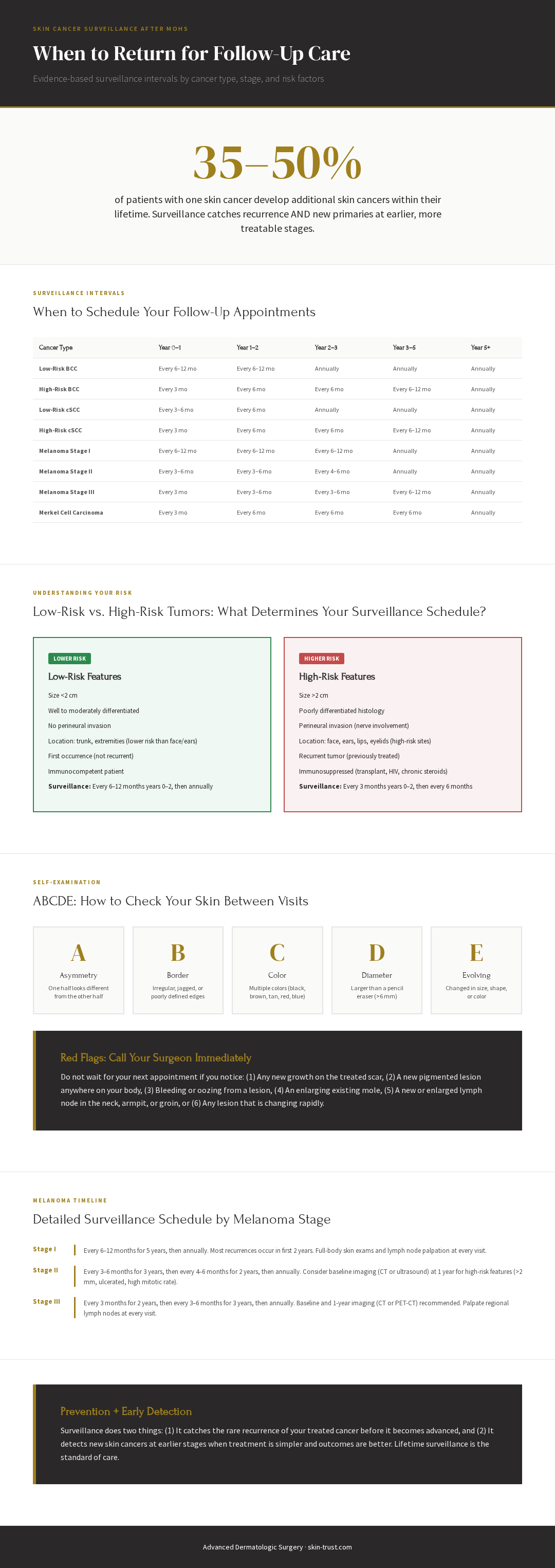
- Surveillance intervals are risk-stratified: BCC every 6–12 months × 2 years then annually; cSCC every 3–6 months for 2 years; melanoma every 3–12 months depending on stage — higher-risk cancers require more frequent visits
- Full-body skin exams for life are standard of care after any skin cancer — this catches new primary tumors early, when they're most treatable
- Between visits, perform monthly self-exams using ABCDE criteria — asymmetry, border irregularity, color variation, diameter >6mm, and evolution (change over time) are warning signs
- Annual sun protection and ongoing skin cancer prevention are essential — the same factors that caused the first cancer will likely produce additional ones if not addressed
Why is surveillance essential after Mohs surgery?
Mohs surgery cures the cancer it is designed to remove—but it does not prevent new skin cancers from developing. Patients with one skin cancer are at significantly elevated lifetime risk for additional skin cancers. Surveillance catches recurrence of the treated cancer AND new primary tumors at earlier, more treatable stages.
Mohs surgery is one of the most effective cancer treatments in dermatology, with cure rates exceeding 99% for the treated lesion. But this high cure rate applies only to the tumor under the microscope. The patient's overall skin cancer risk is not cured by Mohs surgery—it persists.
Here is the critical epidemiology: A patient with one BCC has a 35–50% lifetime risk of developing at least one additional skin cancer. This risk is highest in the first 5 years post-treatment and increases with age, sun exposure, and immunosuppression.
Why is this? Because whatever factors caused the first skin cancer—chronic sun exposure, genetic predisposition, immunosuppression—are still present. A new skin cancer can develop on the same anatomic site (recurrence) or on a completely different site (new primary tumor).
Surveillance accomplishes two things:
It catches recurrence early. Although Mohs cure rates exceed 99%, occasional recurrences do occur (typically within 1–2 years). Early detection allows for prompt re-treatment before the recurrent tumor becomes larger or more invasive.
It catches new primary tumors at earlier stages. A melanoma detected at Stage I has dramatically better prognosis than one detected at Stage III. A cSCC detected as <2 cm has lower metastatic risk than one detected at >2 cm. Early detection transforms treatment options and outcomes.
"The moment I finish removing a skin cancer, I start thinking about what the patient's risk is for another one. Mohs surgery was amazingly successful—the tumor is gone. But now I have a patient who has proven themselves to be a skin cancer patient, and my job is to catch the next one before it becomes advanced. That requires a structured surveillance plan, and it requires the patient to understand why showing up to appointments matters, even if their skin looks fine."
— Thomas L.H. Hocker, M.D., M.Phil.
The spectrum of skin cancer risk after Mohs surgery
Risk of recurrence and new primaries varies dramatically by cancer type, histologic factors, size, location, and patient factors (age, immune status, sun exposure). Low-risk BCCs have recurrence rates <1%, while high-risk melanomas (Stage III) have recurrence rates >30%. Surveillance intervals scale with risk.
Factors that determine surveillance intensity
| Risk Factor Category | High-Risk Features | Surveillance Implication |
|---|---|---|
| Cancer Type | Melanoma >> cSCC >> BCC | Melanoma requires most intensive surveillance |
| Size | >2 cm increases risk | Larger lesions require closer follow-up |
| Histologic Grade | Poor differentiation, perineural invasion | Higher grade = more frequent visits |
| Location | Head/neck, ears, lips (higher risk) vs. trunk/extremities (lower risk) | Location impacts both recurrence and cosmetic concern |
| Patient Age | Younger patients have longer lifetime risk | Lifetime surveillance burden is higher |
| Immunosuppression | Transplant, HIV, chronic immunosuppression | Risk of multiple simultaneous cancers |
| Fitzpatrick Skin Type | Lighter skin types (I–II) have higher risk | Baseline risk is higher |
| Sun Exposure History | Chronic occupational or recreational sun exposure | Future sun exposure will likely continue |
BCC Surveillance: The 6–12 Month Protocol
For low-risk basal cell carcinoma (BCC), surveillance is every 6–12 months for the first 2 years, then annually thereafter. For high-risk BCC (size >2 cm, perineural invasion, poor location for Mohs, immunosuppressed patient), surveillance is every 3–6 months for 2 years, then every 6–12 months for 3 years, then annually. These guidelines are from the 2018 American Academy of Dermatology consensus statement (Kim et al., 2018).
BCC is the most common skin cancer and, fortunately, the lowest-risk malignancy we treat. Recurrence rates are <1% after successful Mohs surgery. However, patients with BCC are at significant risk for new primary BCCs—the same factors that caused the first cancer will likely produce additional cancers over a lifetime.
Low-Risk BCC
Definition: Primary lesion <2 cm, well-differentiated to moderately differentiated, no perineural invasion, location not particularly high-risk (not central face, ear, lid, lip, genitals, or a Mohs micrographic surgery defect >4 mm), and patient is not immunosuppressed.
Surveillance schedule:
- Year 1: Every 6–12 months (2 visits minimum)
- Year 2: Every 6–12 months (1–2 visits)
- Year 3 and beyond: Annually
What to examine: The treated scar site (for recurrence) and full body skin (for new primaries). Dermoscopy is helpful for identifying new suspicious lesions early.
High-Risk BCC
Definition: One or more of the following: lesion >2 cm, poor differentiation, perineural invasion, location on central face/ears/lips/eyelids/genitals, previous BCC recurrence, immunosuppressed status, or failed prior treatment.
Surveillance schedule:
- Year 1: Every 3 months (4 visits minimum)
- Year 2: Every 6 months (2 visits minimum)
- Year 3–5: Every 6–12 months (1–2 visits per year)
- Year 5 and beyond: Annually
What to examine: Close scrutiny of the treated site for any sign of recurrence, full-body skin exam, and lymph node exam in the regional drainage area (though BCC rarely metastasizes).
cSCC Surveillance: The 3–6 Month Protocol
For low-risk cutaneous squamous cell carcinoma (cSCC), surveillance is every 3–6 months for 2 years, then annually. For high-risk cSCC (size >2 cm, poor differentiation, perineural invasion, immunosuppressed, or history of metastasis), surveillance is every 3 months for 2 years, then every 6 months for 3 years, then annually. These guidelines are from the 2018 American Academy of Dermatology consensus statement (Kim et al., 2018).
cSCC carries higher recurrence and metastatic potential than BCC, particularly when high-risk features are present. Surveillance must be more intensive.
Low-Risk cSCC
Definition: Lesion <2 cm, well to moderately differentiated, no perineural invasion, no history of radiation or immunosuppression, and located on low-risk anatomic sites (trunk, extremities).
Surveillance schedule:
- Year 1: Every 3–6 months (2–4 visits minimum)
- Year 2: Every 3–6 months (2 visits minimum)
- Year 3 and beyond: Annually
What to examine:
- Treated scar site (for local recurrence)
- Regional lymph nodes (for metastatic disease)
- Full-body skin exam (for new primaries)
- Dermoscopy of suspicious lesions
High-Risk cSCC
Definition: One or more of the following: lesion >2 cm, poorly differentiated histology, perineural invasion, immunosuppression (post-transplant, HIV, chronic oral steroids), recurrent or previously treated cSCC, or location on high-risk anatomic sites (face, ears, lips, genitals, or areas of chronic inflammation/radiation).
Surveillance schedule:
- Year 1: Every 3 months (4 visits minimum)
- Year 2: Every 6 months (2 visits minimum)
- Year 3–5: Every 6 months (1–2 visits per year)
- Year 5 and beyond: Annually
What to examine:
- Treated site with high scrutiny
- Regional lymph nodes at EVERY visit (palpate neck, submandibular, parotid, preauricular nodes)
- Full-body skin exam
- Consider imaging (CT or MRI of primary site) at 1 year for high-grade tumors or perineural invasion
- Consider imaging of regional lymph nodes (ultrasound or CT) if any suspicious lymphadenopathy
Melanoma Surveillance: Stage-Specific Protocols
Melanoma surveillance is determined by TNM stage and follows strict NCCN/AAD guidelines. Stage I melanoma requires every 6–12 months × 5 years then annually. Stage II requires every 3–6 months × 3 years then annually. Stage III requires every 3 months × 2 years then every 3–6 months × 3 years then annually. These guidelines are from the 2019 American Academy of Dermatology consensus statement (Swetter et al., 2019).
Melanoma is fundamentally different from BCC and cSCC in terms of surveillance intensity and the role of imaging.
Stage I Melanoma (T1a/T1b, N0, M0)
Definition: Tumor thickness ≤1 mm (T1), no regional lymph node involvement, no distant metastases.
Surveillance schedule:
- Years 0–5: Every 6–12 months (1–2 visits per year). Some recent guidelines recommend every 3–6 months × 2 years, then every 6–12 months × 3 years, reflecting the understanding that recurrence risk is highest early.
- Year 5 and beyond: Annually
What to examine:
- Clinical skin exam (treated site and entire skin surface)
- Palpation of regional lymph nodes (most important predictor of recurrence)
- No routine imaging (CT, PET, ultrasound) recommended for Stage I unless patient is symptomatic or you have clinical suspicion of metastasis
Stage II Melanoma (T2–T4, N0, M0)
Definition: Tumor thickness >1 mm, no regional lymph node involvement, no distant metastases. Often divided into IIA (1–2 mm), IIB (2–4 mm), and IIC (>4 mm).
Surveillance schedule:
- Years 0–3: Every 3–6 months (2–4 visits per year)
- Years 3–5: Every 4–6 months (2 visits per year)
- Year 5 and beyond: Annually
What to examine:
- Clinical skin exam (meticulous—look for any new pigmented lesions)
- Palpation of regional lymph nodes at EVERY visit
- Consider baseline lymph node imaging (ultrasound or CT) and repeat imaging at 1 year if high-risk features (poor prognosis, >2 mm thickness)
- Labs: Some physicians order baseline LDH (lactate dehydrogenase) and repeat every 3–6 months if abnormal, though data supporting this is limited
Stage III Melanoma (Any T, N1–N3, M0)
Definition: Tumor of any thickness with regional lymph node metastases but no distant metastases. This is divided into IIIA (1 positive lymph node), IIIB (2–3 positive nodes or in-transit metastases), IIIC (4+ positive nodes), and IIID (any with in-transit/satellite metastases).
Surveillance schedule:
- Years 0–2: Every 3 months (4 visits per year)
- Years 2–5: Every 3–6 months (2–4 visits per year)
- Year 5 and beyond: Annually (or as clinically indicated)
What to examine:
- Clinical skin exam at EVERY visit (in-transit recurrence can appear between primary site and regional nodes)
- Palpation of regional lymph nodes (particularly those draining the primary site)
- Imaging: Baseline CT chest/abdomen/pelvis (or PET-CT) at diagnosis. Repeat imaging at 1 year and as clinically indicated based on risk.
- Labs: LDH at baseline and during surveillance
- Neurologic exam (if brain metastases are suspected)
- Evaluation of lymph node sites for clinical evidence of recurrence
Merkel Cell Carcinoma and Other Cutaneous Malignancies
Merkel cell carcinoma (MCC) is rare but aggressive, with high recurrence and metastatic potential. Surveillance is every 3 months for 2 years, then every 6 months for 3 years, then annually. DFSP (dermatofibrosarcoma protuberans) and AFX (atypical fibroxanthoma) require annual follow-up indefinitely, as late recurrences can occur.
Merkel Cell Carcinoma (MCC)
Definition: Neuroendocrine malignancy with high propensity for recurrence and metastasis. Can present with regional lymph node involvement or distant metastases at diagnosis.
Surveillance schedule:
- Years 0–2: Every 3 months (4 visits per year)
- Years 2–5: Every 6 months (2 visits per year)
- Year 5 and beyond: Annually
What to examine:
- Regional lymph node exam at EVERY visit (MCC metastasizes to lymph nodes in ~25% of cases)
- Skin exam of primary site and draining lymphatic areas
- Baseline imaging (CT or PET-CT) and repeat imaging at 1–2 years per oncology recommendations
- Consider sentinel lymph node biopsy at time of treatment (if not already done)
DFSP (Dermatofibrosarcoma Protuberans)
Surveillance schedule:
- Annually for life (DFSP can recur many years later, so lifetime surveillance is prudent)
What to examine:
- Clinical exam of primary site (look for any nodules or new growth)
- Feel for any recurrence at the original scar site
AFX (Atypical Fibroxanthoma)
Surveillance schedule:
- Annually for 5 years, then consider surveillance based on risk (AFX is generally lower-risk than other cutaneous malignancies, but late recurrence is possible)
Comprehensive Skin Cancer Surveillance Table
| Cancer Type | Year 0–1 | Year 1–2 | Year 2–3 | Year 3–5 | Year 5+ |
|---|---|---|---|---|---|
| Low-Risk BCC | Every 6–12 mo | Every 6–12 mo | Annually | Annually | Annually |
| High-Risk BCC | Every 3 mo | Every 6 mo | Every 6 mo | Every 6–12 mo | Annually |
| Low-Risk cSCC | Every 3–6 mo | Every 6 mo | Annually | Annually | Annually |
| High-Risk cSCC | Every 3 mo | Every 6 mo | Every 6 mo | Every 6–12 mo | Annually |
| Melanoma Stage I | Every 6–12 mo | Every 6–12 mo | Every 6–12 mo | Annually | Annually |
| Melanoma Stage II | Every 3–6 mo | Every 3–6 mo | Every 4–6 mo | Annually | Annually |
| Melanoma Stage III | Every 3 mo | Every 3–6 mo | Every 3–6 mo | Every 6–12 mo | Annually |
| Merkel Cell Carcinoma | Every 3 mo | Every 6 mo | Every 6 mo | Every 6 mo | Annually |
| DFSP | Annually | Annually | Annually | Annually | Annually |
| AFX | Annually | Annually | Annually | Annually | Consider per risk |
Data sourced from peer-reviewed, PubMed-indexed publications
What to look for between surveillance visits
Between scheduled surveillance appointments, patients should perform self-examination using ABCDE criteria (for melanoma) and watch for any new growths, changes to the treated scar, or enlarged lymph nodes. Any concerning finding should prompt an unscheduled visit rather than waiting for the next appointment.
ABCDE Criteria for Melanoma Self-Exam
Even patients treated for BCC or cSCC should learn these criteria, because the same sun-damaged skin that produced one cancer can produce any type of skin cancer.
- Asymmetry: Is one half of the lesion different from the other half? Melanomas are typically asymmetric.
- Border: Is the border irregular, jagged, or poorly defined? Benign nevi have smooth, well-demarcated borders.
- Color: Does the lesion have multiple colors (black, brown, tan, red, blue)? Melanomas often have color variation.
- Diameter: Is the lesion >6 mm (the size of a pencil eraser)? Melanomas are often >6 mm at diagnosis, though small melanomas do exist.
- Evolving: Has the lesion changed in size, shape, or color in recent weeks to months? Any change is concerning.
Additional Red Flags Between Visits
Patients should alert their surgeon immediately if they notice:
New growth on treated scar site: Any bumpy, scaly, or nodular growth on the surgical scar itself could represent recurrence.
New pigmented lesion anywhere on body: Especially if it is asymmetric, has irregular borders, or has multiple colors.
Bleeding or oozing lesion: Spontaneous bleeding from a skin lesion is concerning for malignancy.
Enlarging existing mole: Any mole that grows over weeks to months warrants evaluation.
Enlarged lymph node: A new or enlarging lymph node in the regional drainage area (neck, armpit, groin) can indicate metastasis.
Non-healing wound or ulcer: Persistent non-healing wounds can be cSCC or other malignancies.
Itching or pain in a lesion: Although benign lesions can itch, persistent symptoms should prompt evaluation.
The role of dermoscopy in surveillance
Dermoscopy (dermoscopic magnification, typically 10x) significantly improves the diagnostic accuracy of skin exams. For melanoma detection, dermoscopy improves sensitivity (ability to detect melanoma) from ~80% to 90–95% and specificity (ability to exclude benign lesions) from ~80% to 85–90%. For surveillance visits, dermoscopy of questionable lesions should be standard.
Dermoscopy is a magnified view of the skin using specialized illumination. It is not a biopsy—it does not require tissue sampling. But it reveals details of color, pattern, and structure that are invisible to the naked eye.
Why it matters for surveillance: In a patient with prior skin cancer (especially melanoma), the baseline skin often has multiple atypical nevi that can mimic melanoma. Dermoscopy helps distinguish benign atypical nevi from melanoma based on specific patterns (ABCD rule of dermoscopy, Menzies criteria, etc.).
For an experienced dermatologist, dermoscopy is one of the most useful tools in the surveillance examination. Any lesion that is questionable on clinical exam should be examined with dermoscopy before deciding whether biopsy is needed.
Imaging and laboratory testing in surveillance
Imaging (CT, PET, MRI) and lab testing (LDH) are not routinely recommended for Stage I melanoma or BCC/cSCC surveillance in asymptomatic patients. For Stage II melanoma, baseline imaging is reasonable, with repeat at 1 year for high-risk features. For Stage III melanoma, imaging is recommended. For high-risk cSCC, baseline and 1-year imaging of regional nodes is appropriate. These guidelines balance detecting metastatic disease against avoiding unnecessary testing and false positives.
When Imaging is Indicated
| Cancer Type | Indication for Imaging |
|---|---|
| Stage I Melanoma | Only if symptomatic or clinical suspicion of metastasis |
| Stage II Melanoma | Consider baseline ultrasound or CT; repeat at 1 year if high-risk features (>2 mm, ulcerated, high mitotic rate) |
| Stage III Melanoma | Baseline CT chest/abdomen/pelvis or PET-CT; repeat at 1 year and per oncology recommendations |
| High-Risk cSCC | Consider ultrasound or CT of regional lymph nodes at baseline and 1 year |
| Low-Risk BCC/cSCC | No routine imaging unless symptomatic |
| Merkel Cell Carcinoma | Baseline CT or PET-CT; repeat per oncology recommendations |
LDH (Lactate Dehydrogenase) Testing
LDH is a non-specific marker that can be elevated in melanoma, particularly Stage III or IV disease. Some physicians obtain baseline LDH and repeat every 3–6 months, though the evidence supporting this is limited. LDH is not specific (can be elevated for many reasons) and not sensitive (can be normal even with metastatic disease). Its role in asymptomatic surveillance remains controversial.
FAQ: Skin Cancer Surveillance
Q: Do I really need surveillance if my cancer was completely removed? A: Yes. Although Mohs surgery has cure rates >99% for the treated lesion, you remain at significant risk for new skin cancers. The same factors that caused the first cancer (sun exposure, genetic predisposition) are still present. Surveillance catches recurrence early (before it becomes advanced) and new primaries at earlier, more treatable stages.
Q: How long do I need surveillance? A: Indefinitely, though intensity decreases over time. For most patients, the highest-risk period is the first 2–5 years. But skin cancer can recur many years later. A reasonable approach is annual full-body skin exams for life, with more frequent visits in the first 2–5 years.
Q: Can I do self-exams instead of coming to the office? A: Self-exams are important and supplement (not replace) professional surveillance. Self-exams allow you to monitor for obvious changes between visits. But professional exams with dermoscopy are more sensitive at detecting early melanoma, cSCC, and new primaries. A typical surveillance strategy combines both: patient self-exam every month + professional exams every 3–12 months depending on risk.
Q: What should I do if I find a new bump or growth on my skin? A: Call your surgeon immediately. Do not wait for your next scheduled appointment. If it is a benign lesion, the visit provides reassurance. If it is concerning, early detection is critical. Many skin cancers detected clinically (as opposed to incidentally) are caught because the patient noticed something and reported it.
Q: Do I need imaging (CT, PET scan) after treatment? A: Not routinely for Stage I BCC/cSCC or Stage I melanoma unless you have symptoms or your surgeon suspects metastatic disease. For Stage II melanoma and high-risk cSCC, imaging at 1 year is reasonable. For Stage III melanoma, imaging is recommended. Discuss with your surgeon what imaging, if any, is appropriate for your specific situation.
Q: What if my surveillance visits show no new cancers—can I stop coming? A: No. The absence of cancer in the first 2 years does not mean cancer will not develop later. Recurrence and new primaries can develop years after treatment. Ongoing surveillance is necessary, though intervals may extend (e.g., annual visits instead of every 3 months after year 5).
Q: Should I see a dermatologist or my primary care doctor for surveillance? A: Ideally, surveillance by the dermatologist or Mohs surgeon who treated you is preferred because they know your history, can compare to baseline, and can perform dermoscopy and advanced biopsies if needed. A dermatologist is trained in skin cancer recognition and has the tools (dermoscopy, biopsy equipment) to evaluate concerning lesions efficiently. Annual surveillance by your Mohs surgeon is ideal; if cost or distance is a barrier, at minimum annual full-body skin exams by a dermatologist are recommended.
Q: What is the risk that my treated cancer will come back? A: Recurrence risk depends on cancer type and characteristics. For low-risk BCC, <1%. For high-risk BCC, 1–5%. For low-risk cSCC, 1–3%. For high-risk cSCC, 5–15%. For Stage I melanoma, 1–5%. For Stage II melanoma, 10–20%. For Stage III melanoma, >30%. These are rough estimates; your surgeon can provide personalized risk based on your pathology.
Q: How often do patients develop a new skin cancer after Mohs surgery? A: Approximately 35–50% of patients with one skin cancer develop at least one additional skin cancer within their lifetime. The risk is highest in the first 5 years and decreases thereafter, but additional cancers can develop decades later. This is why lifetime surveillance is recommended.
Q: What should I do to reduce my risk of another skin cancer? A: Sun protection (mineral sunscreen SPF 30+, protective clothing, hats, shade-seeking), avoid tanning beds, and skin self-exams every month. Your surgeon will provide specific sun protection recommendations based on your wound healing requirements.
Q: Can you tell if a lesion is cancer just by looking at it without a biopsy? A: No. While experienced dermatologists can identify many cancers based on clinical appearance, some malignancies look deceptively benign, and some benign lesions look concerning. Dermoscopy improves accuracy but is not definitive. When there is any clinical suspicion, biopsy is the only way to confirm diagnosis. If your surgeon recommends a biopsy, it is appropriate to proceed.
Q: What is a sentinel lymph node biopsy, and should I have one? A: A sentinel lymph node biopsy (SLNB) involves injecting dye and/or radioactive tracer near the primary tumor, identifying the first lymph node(s) to which cancer would typically drain, and biopsying that node to check for metastatic disease. SLNB is recommended for melanoma >0.8–1 mm thick or with other high-risk features (ulceration, high mitotic rate). It is not routinely indicated for BCC or cSCC unless there is clinical evidence of lymph node involvement. Your surgeon or oncologist can discuss whether SLNB is appropriate for your specific tumor.
References
Kim JY, Kozlow JH, Miteva M, Olbricht SM, Rigel DS. Guidelines of care for basal cell carcinoma. Journal of the American Academy of Dermatology. 2018;78(3):540-559. PMID: 29331385
Kim JYS, Kozlow JH, Miteva M, Olbricht SM, Rigel DS. Guidelines of care for cutaneous squamous cell carcinoma. Journal of the American Academy of Dermatology. 2018;78(3):560-578. PMID: 29331386
Swetter SM, Tsao H, Bichakjian CK, et al. Guidelines of care for the management of melanoma: Surveillance of patients and follow-up assessment. Journal of the American Academy of Dermatology. 2019;80(2):308-318. PMID: 30392755
Medical Disclaimer
This article is provided for educational purposes only and does not constitute medical advice. The information presented is based on peer-reviewed, published research and clinical practice guidelines from the American Academy of Dermatology, National Comprehensive Cancer Network (NCCN), and American College of Mohs Surgery. However, individual patient factors, tumor characteristics, and risk profiles vary significantly.
Before establishing a surveillance plan, consult with your treating surgeon to confirm recommendations are appropriate for your specific tumor type, stage, and personal risk factors. Do not discontinue or modify medical recommendations provided by your oncologist or surgeon without explicit approval.
Treatment decisions should be based on individual patient factors, not general guidelines alone. Seek immediate medical attention if you notice any new skin growth, changes to a treated area, or enlarged lymph nodes.
