
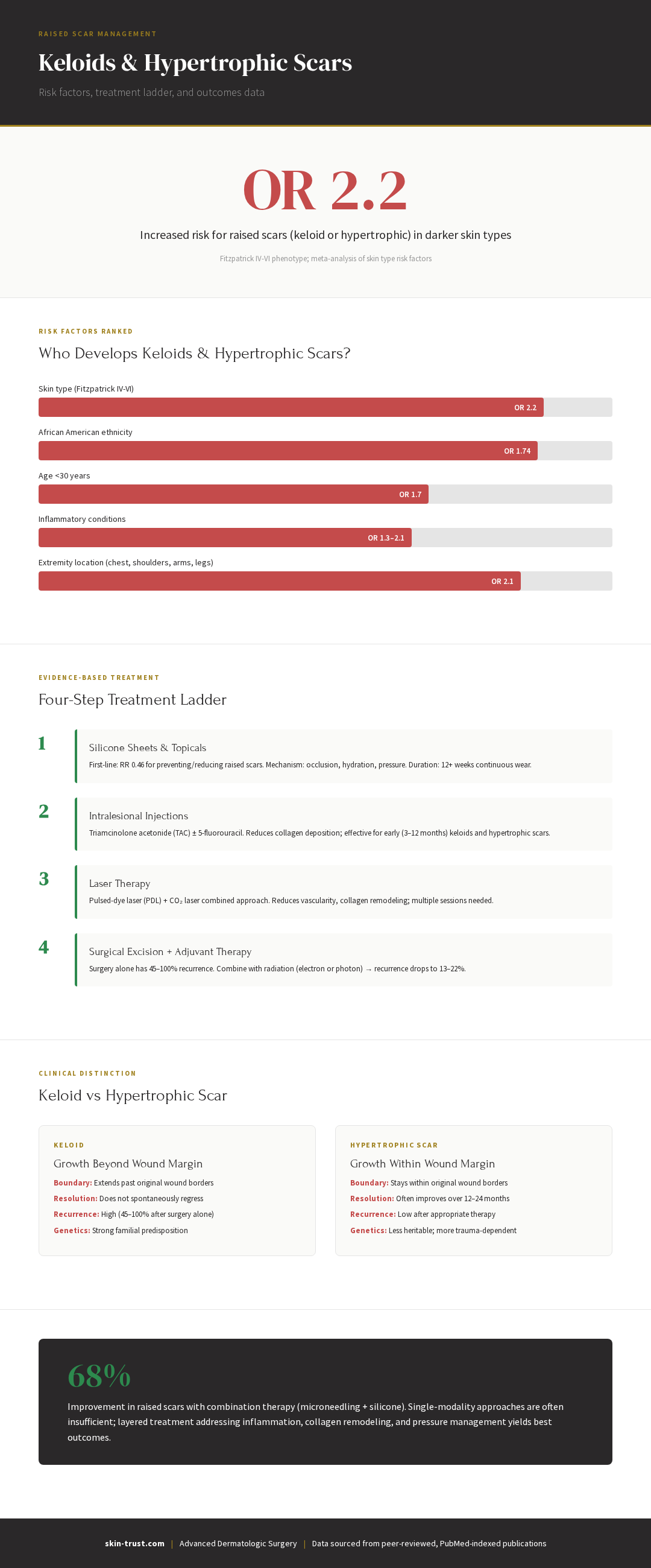
- Keloids extend beyond original wound borders; hypertrophic scars stay within boundaries — the distinction determines treatment urgency (keloids require intervention; hypertrophic scars often improve alone)
- High-risk patients: age <30, Fitzpatrick IV–VI skin, Black/African American ancestry, and high-tension anatomic areas (chest, shoulders, back, earlobes) — these patients need preventive measures and closer surveillance
- Silicone gel/sheets reduce both keloid and hypertrophic scar formation (RR 0.46) — start 2 weeks post-op and continue ≥12 weeks minimum; benefit extends to all skin tones
- Tension-offloading with paper tape prevents hypertrophic scarring entirely in some studies (41% → 0%) — critical for high-risk patients and high-tension body areas
- Intralesional triamcinolone (TAC) 10–40 mg/mL every 2–4 weeks is first-line for established keloids — multiple injections are typically needed; 5-FU can be added for treatment-resistant cases
- Surgical excision plus adjuvant radiation reduces keloid recurrence from 45–100% down to 13–22% — optimal for large, recurrent, or treatment-resistant keloids
Evidence Snapshot
Keloids and hypertrophic scars are a common, disfiguring consequence of surgery—especially in darker skin tones and certain body locations. They're not just a cosmetic problem: they cause itching, pain, functional limitation (if over a joint), and significant psychological distress. Yet many patients don't know they're at high risk until after a scar forms. And many dermatologists don't have a systematic approach to treatment. This article walks you through who's at risk, how to prevent problematic scars, and the evidence-based treatment ladder when scars do form.
Part 1: What's the Difference Between Hypertrophic Scars and Keloids?
Hypertrophic Scars
Definition: Raised, firm, red scars that stay within the boundaries of the original wound. Think of them as the scar tissue trying to overcompensate to repair the wound.
Characteristics:
- Elevated above the skin surface
- Red or pink (vascular, still remodeling)
- Firm to palpation
- Itching, tenderness common
- Confined to original wound borders
- Usually improve over time (12–24 months) as collagen remodels and vascular activity decreases
Timeline: Appear weeks to months post-op; may improve spontaneously over 1–2 years without treatment.
When it happens: Hypertrophic scars are common after:
- High-tension closures
- Wounds over joints (which move a lot)
- Wounds on the anterior chest
- Wounds in darker skin tones (though not exclusively)
Keloids
Definition: Raised scars that extend beyond the original wound borders. Keloids are the scar tissue growing aggressively into normal surrounding skin.
Characteristics:
- Elevated above skin surface
- Extend beyond original wound margins
- Red, pink, or purple
- Firm, sometimes painful or itchy
- Can be quite large (1–2cm or larger)
- Do not regress spontaneously without treatment
- Recur frequently after simple excision (45–100% recurrence without adjuvant therapy)
Timeline: Appear weeks to months post-op; worsen over time without treatment.
When it happens: Keloids are more common in:
- Fitzpatrick IV–VI skin (3–15× higher risk than Fitzpatrick I–III)
- Black/African American patients (10–15% lifetime risk)
- Age <30
- High-tension areas (anterior chest, shoulders, earlobes, back)
- Family history of keloid formation
Key Distinction
| Feature | Hypertrophic Scar | Keloid |
|---|---|---|
| Extends beyond wound? | No (stays within borders) | Yes (grows beyond original wound) |
| Spontaneous regression? | Yes (often over 12–24 months) | No (persists indefinitely) |
| Ethnicity risk | Modest increase in Fitzpatrick IV–VI | High increase in Fitzpatrick IV–VI |
| Treatment needed? | Optional; may improve alone | Usually needed; won't resolve alone |
| Recurrence after excision | Low (5–10%) | High (45–100% without adjuvant therapy) |
Data sourced from peer-reviewed, PubMed-indexed publications
Part 2: Risk Factors for Keloid and Hypertrophic Scar Formation
Genetics and Ethnicity
Meta-analysis (Sharma et al., 2025): Risk factors for keloid formation.
Fitzpatrick Skin Type:
- Fitzpatrick IV–VI (darker skin): OR 2.2 (2.2× higher risk than Fitzpatrick I–III)
- The risk is highest in Black, Hispanic, and Asian populations
Ancestry:
- Black/African American: OR 1.74
- Lifetime risk: 10–15% in Black patients vs. 0.1% in White patients
Why the difference? Darker skin tones have:
- More active melanocytes and inflammatory cells
- Genetic predisposition to altered collagen remodeling
- Possible differences in transforming growth factor-β (TGF-β) signaling, which drives fibroblast proliferation
Age
Age <30: OR 1.7 (younger patients have higher risk)
Why? Younger skin has:
- More active collagen remodeling
- Higher growth factor levels
- Greater inflammatory response
Inflammatory Comorbidities
Conditions that increase keloid/hypertrophic scar risk:
- Acne (OR 1.3–2.1): Inflammatory cascade triggers fibroblast proliferation
- Atopic dermatitis/eczema (OR 1.5–2.0): Chronic inflammation primes the scar response
- Psoriasis: Chronic inflammatory state increases scar tissue formation
- Systemic lupus erythematosus (SLE): Autoimmune inflammation
Implication: If you have acne or eczema and darker skin, your keloid risk is compounded.
Anatomic Location (High-Tension Areas)
Highest risk locations:
- Anterior chest: 10–15× more likely to form keloids than other body sites
- Shoulders: High-tension area; frequently affected
- Upper back: Especially in men (deltoid region)
- Earlobes: Piercing or surgical trauma commonly triggers keloids
- Upper arms: Especially over the biceps
- Joints (shoulders, elbows, knees): Movement and tension perpetuate scar growth
Lower risk locations:
- Face (except jawline)
- Scalp
- Lower legs
- Palms and soles
Why location matters: High-tension areas have:
- Continuous muscle movement (stretching the scar)
- Thick skin with high collagen density
- Rich vascular supply
- Pressure and friction (if over a joint)
Wound Characteristics
High-risk wound features:
- Large defects: Larger wounds require more collagen; bigger scars result
- High-tension closures: Tension perpetuates scar tissue formation
- Wounds requiring flaps: More extensive surgery → more inflammation → higher scar risk
- Delayed healing: Prolonged inflammation increases keloid risk
- Infection: Infection increases inflammatory response and scarring
Lower-risk features:
- Small wounds: <5mm defects rarely keloid
- Low-tension closures: Tension-free closures minimize scarring
- Primary healing: Wounds that heal without complications scar better
Arterial Inflammation and Vascular Changes
Emerging research (Sugimoto et al., 2022, PMID 34537794): Arteriosclerosis and keloid formation. Study suggests that arterial inflammation and vascular changes may contribute to keloid pathophysiology.
Implication: Patients with cardiovascular risk factors or chronic vascular disease may have increased keloid risk, though the mechanism is still being elucidated.
Part 3: Prevention — What to Do Before and After Surgery
Pre-Operative Risk Stratification
High-risk patient profile:
- Fitzpatrick IV–VI skin
- Age <30
- Personal or family history of keloids
- Anterior chest or shoulder defect location
- Inflammatory comorbidities (acne, eczema, SLE)
If you're high-risk, tell your surgeon. They can:
- Optimize surgical technique (minimal tension, RSTL alignment, flap use if needed)
- Plan preventive post-op therapy (silicone starting week 1, pressure garment if anterior chest)
- Consider different wound closure options (flap instead of primary closure)
- Discuss intralesional steroid prophylaxis if extremely high-risk
Immediate Post-Op Prevention (Weeks 1–6)
Paper Tape (Week 1–6)
Evidence: (Atkinson et al., 2005), OR 13.6 reduction in hypertrophic scars.
Apply: Micropore or elastic adhesive tape perpendicular to the wound for 6 weeks.
Benefit: Mechanical tension offloading; essential in the critical healing window.
Silicone Gel Sheets or Cream (Week 2–12+)
Evidence: (Wang et al., 2020), meta-analysis of multiple RCTs. Relative risk (RR) 0.46 for hypertrophic scarring. Cochrane review: silicone significantly reduces risk of hypertrophic scars.
How to use:
- Start week 2–3 (once wound is fully closed)
- Wear 12+ hours daily for 8–12 weeks
- For high-risk patients: continue for 3–6 months
Effect: Occlusion and hydration; reduces redness and firmness; flattens raised scars.
Cost: $20–100 for 2–3 months.
Pressure Therapy (High-Risk Patients, Anterior Chest/Shoulders)
Evidence: Meta-analysis of 12 RCTs, 710 patients. Pressure therapy (garments, elastic bandages) achieves Vancouver Scar Scale (VSS) mean difference −0.58, indicating modest improvement in scar appearance.
For anterior chest/shoulders: Custom pressure garments (Jobst, Comprilan) worn 23 hours daily for 3–6 months.
Cost: $200–2,000 for custom garments.
Who needs it: Patients with high-risk location (anterior chest), high-risk ethnicity (Fitzpatrick IV–VI), and personal or family history of keloids.
Massage (Weeks 2–6+)
Technique: Firm, directional massage perpendicular to the scar, 2–3 times daily for 3–5 minutes.
Mechanism: Remodels collagen, improves circulation, reduces firmness.
Evidence: Modest benefit for symptom relief (itch, firmness) and cosmetic appearance. Works best combined with silicone and pressure therapy.
High-Risk Preventive Strategy: Combination Approach
For a patient at very high risk (Fitzpatrick V skin, anterior chest, age 22, family history of keloids):
Weeks 1–6: Paper tape + gentle wound care
Weeks 2–12: Silicone gel sheets (12+ hours daily) + pressure garment (23 hours daily) + massage (2–3 times daily)
Week 4–6: If any signs of hypertrophic scar or keloid formation, consider early intralesional TAC injection (prophylactic)
Months 3–6: Continue silicone and pressure as scar matures
Result: Combined approach reduces hypertrophic scar formation and prevents early keloid formation better than any single modality.
Part 4: Treatment Ladder for Hypertrophic Scars and Keloids
Level 1: Silicone and Pressure Therapy (First-Line Conservative)
Timing: Hypertrophic scars detected at 2–4 months post-op.
What to do:
- Silicone gel sheets 12+ hours daily for 8–12 weeks
- Pressure garment (if anterior chest/shoulders) 23 hours daily for 3–6 months
- Massage 2–3 times daily
Success rate: 40–60% of hypertrophic scars improve significantly (flatter, less red, less firm) with silicone and pressure alone.
Timeline: 8–12 weeks to see improvement; continued use for 3–6 months for optimal benefit.
Cost: $100–300 total.
Level 2: Intralesional Injections (For Persistent Hypertrophic Scars or Early Keloids)
Intralesional Triamcinolone (TAC)
What it is: Corticosteroid injection directly into the scar tissue.
How it works: TAC suppresses inflammatory cytokines and fibroblast proliferation, causing scar tissue to shrink and flatten.
Dosing: 10–40 mg/mL (concentration and volume vary by scar size and depth). Injected intradermally and subdermally.
Results:
- 70–90% of hypertrophic scars improve with TAC
- Flattening visible within 2–4 weeks
- Redness decreases over weeks
- Firm scar tissue softens
Timeline: 3–6 injections spaced 4–6 weeks apart for optimal effect.
Cost: $100–300 per injection session; not always covered by insurance.
Complications: Telangiectasia (small blood vessels), skin atrophy (depression), depigmentation (especially in darker skin). These are usually temporary and resolve over weeks to months.
Intralesional TAC + 5-Fluorouracil (5-FU)
What it is: TAC combined with 5-FU (chemotherapy agent that inhibits fibroblast proliferation).
Why combine them: 5-FU enhances TAC's anti-fibroblast effect, improving outcomes over TAC alone for keloids.
Results: Superior to TAC monotherapy for keloid recurrence reduction.
Dosing: Typically 40 mg/mL TAC mixed with 50 mg/mL 5-FU in a 1:1 ratio.
Timeline: 4–6 injections spaced 3–4 weeks apart.
Cost: $200–500 per injection (slightly more than TAC alone due to 5-FU).
Best for: Keloids (especially recurrent keloids after excision) and severe hypertrophic scars.
Complications: Similar to TAC; 5-FU adds minimal extra risk but may increase post-injection inflammation.
Level 3: Laser Treatment (For Resistant Hypertrophic Scars or Keloids)
Pulsed-Dye Laser (PDL)
What it is: Yellow laser (585–595 nm wavelength) that targets hemoglobin in blood vessels.
How it works: Reduces vascularity (redness) and inflammation in the scar.
Results:
- Reduces redness and erythema
- Modest flattening of raised scars
- Works best when scar is still vascular and red
Timeline: 4–6 treatments spaced 4–6 weeks apart.
Cost: $300–800 per session; 4–6 sessions = $1,200–4,800.
Best for: Hypertrophic scars with significant redness; less effective for pale, mature keloids.
Fractional CO₂ Laser
What it is: Ablative laser that vaporizes columns of scar tissue, stimulating collagen remodeling.
How it works: Creates controlled injury, triggering wound healing and collagen restructuring.
Results:
- 40–70% improvement in scar appearance (flattening, narrowing)
- Can combine with TCA cross for icepick-type keloids
- Redness decreases over months
Timeline: 1–3 treatments spaced 4–8 weeks apart.
Cost: $500–1,500 per session.
Downtime: 5–7 days of redness and crusting; requires sun protection.
Best for: Hypertrophic scars; keloids if combined with intralesional therapy.
Microneedling + Silicone Combination
Study: Microneedling followed by silicone gel application.
Results: 68% improvement in scar appearance (flattening, texture) vs. 47% with silicone alone.
Mechanism: Microneedling disrupts collagen organization and triggers remodeling; silicone provides occlusion and hydration during the healing response.
Cost: $200–500 per microneedling session; combine with silicone ($20–100).
Timeline: 4–6 microneedling sessions spaced 4–6 weeks apart, each followed by silicone for 2–4 weeks.
Level 4: Surgical Excision ± Adjuvant Radiation (For Recurrent or Severe Keloids)
The Problem with Simple Excision
If you excise a keloid and just sew it up (primary closure), the keloid often recurs (45–100% recurrence rate in some studies). Why? Because the trauma of surgery itself can trigger keloid re-formation in genetically predisposed patients.
Excision + Intralesional TAC ± 5-FU
Strategy: Excise the keloid surgically, then immediately inject TAC/5-FU into the wound edges and surrounding tissue.
Results: Reduces recurrence to 20–30%.
Timeline: Excision, immediate post-op injection, then serial injections at 4-6 week intervals.
Best for: Keloids that are painful, limiting function, or have failed conservative treatment.
Excision + Adjuvant Radiation
Strategy: Excise the keloid surgically, then deliver external beam radiation (typically 15–20 Gy in 3–5 fractions) within 24 hours of excision.
Results: Keloid recurrence drops to 13–22% compared to 45–100% without radiation.
How it works: Radiation inhibits fibroblast proliferation and collagen deposition, preventing keloid re-formation.
Timing: Radiation must start within 24 hours of excision for optimal effect.
Who does this: Coordination with a radiation oncologist is needed. coordinates with radiation oncology for patients who need this approach.
Cost: Surgical excision ($1,000–3,000) + radiation ($2,000–5,000), often partially covered by insurance.
Best for: Large, recurrent, or functionally limiting keloids where simple excision has failed.
Part 5: Comparison Table — Treatment Options and Evidence
| Treatment | Evidence Level | Effect Size | Cost | Timeline | Best For |
|---|---|---|---|---|---|
| Silicone gel/sheets | Strong (RCT, meta-analysis) | RR 0.46 for hypertrophic scars | $20–100 | 8–12 weeks | Hypertrophic scars, prevention |
| Pressure garments | Moderate | VSS −0.58 (modest improvement) | $200–2,000 | 3–6 months | High-risk locations (anterior chest/shoulders) |
| Intralesional TAC | Strong | 70–90% improvement | $100–300/session | 3–6 sessions, 4–6 weeks apart | Hypertrophic scars, early keloids |
| Intralesional TAC + 5-FU | Strong | Superior to TAC alone for keloids | $200–500/session | 4–6 sessions | Keloids, severe hypertrophic scars |
| Pulsed-dye laser (PDL) | Moderate | Reduces redness; modest flattening | $300–800/session | 4–6 sessions | Hypertrophic scars with redness |
| Fractional CO₂ laser | Moderate to Strong | 40–70% improvement | $500–1,500/session | 1–3 sessions | Hypertrophic scars; keloids if combined with injections |
| Microneedling | Moderate | 30–60% improvement; 68% + silicone | $200–500/session | 4–6 sessions | Atrophic scars, hypertrophic scars |
| Microneedling + silicone | Moderate to Strong | 68% improvement | $200–600/session + silicone | 4–6 sessions + 2–4 weeks silicone each | Hypertrophic scars, atrophic scars |
| Surgical excision ± TAC/5-FU | Strong | 20–30% recurrence | $1,000–3,000 | 1 procedure + serial injections | Keloids, hypertrophic scars |
| Surgical excision ± radiation | Strong | 13–22% recurrence | $3,000–8,000 | 1 procedure + 3–5 radiation sessions | Large, recurrent, or functionally limiting keloids |
| Massage | Weak to Moderate | Symptom relief, modest cosmetic improvement | $0 | 2–3 times daily, 3–6 months | Adjunct therapy; prevents keloid formation |
Part 6: ADS Approach to Keloids and Hypertrophic Scars
Evaluation
- Distinguish keloid from hypertrophic scar: Clinical exam; biopsy if unclear (helps rule out recurrent skin cancer in scar)
- Assess risk factors: Ethnicity, location, age, inflammatory history, family history
- Estimate maturity: Scar age influences treatment (immature scars may improve spontaneously; mature scars need intervention)
- Functional assessment: Is the scar painful, itchy, limiting movement?
Prevention Strategy
For high-risk patients (before or after surgery):
- Paper tape weeks 1–6
- Silicone weeks 2–12+
- Pressure garment (if anterior chest/shoulders) for 3–6 months
- Massage daily
- Sun protection (UV can darken and worsen scars)
Treatment Algorithm
Hypertrophic scar, 2–6 months old, not responding to silicone: → Intralesional TAC (1–3 sessions)
Persistent hypertrophic scar after TAC: → Fractional laser + continued silicone
Early keloid, <6 months: → Intralesional TAC or TAC + 5-FU (4–6 sessions)
Keloid, >6 months, failed conservative treatment: → Surgical excision + intralesional TAC/5-FU ± adjuvant radiation
Pathology Advantage
Because I'm board-certified in dermatopathology, I can biopsy the scar to:
- Confirm it's a keloid/hypertrophic scar and not a recurrent skin cancer
- Assess depth and extent of collagen
- Evaluate for inflammatory markers that predict treatment response
Radiation Coordination
For patients needing excision + adjuvant radiation, coordinates with a radiation oncologist to ensure:
- Excision is optimized for low tension
- Radiation starts within 24 hours of excision
- Follow-up ensures recurrence is detected early
Part 7: Frequently Asked Questions
Q: I have dark skin and just had a mole removed. I'm terrified I'll develop a keloid. What should I do?
A: Tell your surgeon you're concerned about keloid formation. They can optimize your surgical approach and plan immediate post-op prevention:
- Paper tape for weeks 1–6
- Silicone sheets or cream from week 2–3 onward
- Consider a pressure garment if the defect is on your anterior chest or shoulders
- Daily massage
- Sun protection
If your scar looks hypertrophic or starts forming a keloid at 2–4 months, contact your dermatologist immediately for early intralesional steroid injection. Early intervention prevents progression.
Q: Can I prevent keloids if I have a family history?
A: You can't prevent them entirely if you have genetic predisposition, but you can reduce risk:
- Avoid elective surgery on high-risk areas (anterior chest, shoulders, ears) if possible
- If surgery is necessary, use a dermatologic surgeon experienced with keloid-prone patients
- Start preventive silicone and pressure therapy immediately post-op
- Consider prophylactic intralesional steroid injection at 4–6 weeks if early hypertrophic changes appear
Q: I developed a keloid after ear piercing. Should I have it removed?
A: Keloids on the earlobe are common and often cause cosmetic and functional problems (large keloids can distort the ear). Options:
- Intralesional TAC/5-FU: 4–6 injections, often effective for earlobe keloids
- Surgical excision + TAC/5-FU: If large or failed injections
- Surgical excision ± radiation: If recurrent after excision
Talk with a dermatologist about which approach is best for your specific keloid.
Q: Can hypertrophic scars turn into keloids?
A: Hypertrophic scars and keloids are different entities. A hypertrophic scar may worsen over time (particularly if you keep picking at it or if it's in a high-tension area), but it won't spontaneously transform into a keloid. However, they can coexist: a scar can have both hypertrophic (firm, raised, confined to borders) and keloid (extending beyond borders) features. Proper classification by a dermatologist matters because treatment differs.
Q: I've heard that keloid surgery always fails. Is that true?
A: Simple excision alone has 45–100% recurrence. However, excision combined with adjuvant therapy (intralesional injections or radiation) significantly reduces recurrence:
- Excision + TAC/5-FU: 20–30% recurrence
- Excision + radiation: 13–22% recurrence
So no, keloid surgery doesn't always fail—but you need more than just excision. Modern multimodal therapy is much more effective than older simple excision approaches.
Q: How long do I need to use silicone after surgery?
A: For prevention in high-risk patients:
- Minimum 8–12 weeks continuously
- Better: 3–6 months (until scar matures)
- Even better: Continue indefinitely during periods of high re-injury risk (e.g., if you're prone to scratching or if the scar is in a high-tension area)
For treatment of existing hypertrophic scars:
- 8–12 weeks minimum
- Often 3–6 months for continued improvement
There's no harm in using silicone longer; stop when the scar matures and you're satisfied with appearance.
Q: I'm Black and had a surgical scar that developed into a keloid. Am I destined to keloid with every surgery?
A: Not necessarily, but yes, your risk is higher. A personal history of keloid formation significantly increases risk with future surgeries. However:
- Future scars may not keloid if they're in low-risk locations (face, lower legs, palms/soles)
- Proper surgical technique (low-tension closure, RSTL alignment) reduces risk
- Immediate post-op prevention (silicone, pressure, possibly prophylactic injections) reduces risk
- Not all future scars will keloid
Discuss your keloid history with any surgeon doing elective surgery. They can help minimize risk by choosing location, technique, and preventive approach carefully.
Q: Can I use over-the-counter scar products to prevent keloids?
A: Most over-the-counter products (Mederma, Bio-Oil, etc.) lack strong evidence and don't prevent keloids. Silicone is the only over-the-counter product with robust evidence for preventing hypertrophic scars and supporting keloid prevention.
For actual keloid prevention or treatment, you need:
- Prescription-strength silicone or dermatologist-prescribed products
- Professional pressure garments
- Intralesional steroid injections (prescription, dermatologist-administered)
Don't rely on drugstore creams for serious keloid risk. See a dermatologist.
Q: I have a keloid I've had for years. Is it too late to treat it?
A: No. Mature keloids respond well to:
- Intralesional TAC/5-FU injections (even 10+ year old keloids can flatten significantly)
- Combination therapy (injections + laser)
- Surgical excision + adjuvant therapy
Old keloids are less likely to regress spontaneously, so professional treatment is warranted. Discuss options with a dermatologist.
Part 8: Medical Disclaimer
This article is educational and does not replace professional medical advice. Keloid and hypertrophic scar treatment should be individualized based on scar type, location, depth, patient ethnicity and age, and patient goals. Some treatments (intralesional injections, laser, radiation, surgery) require evaluation and treatment by a board-certified dermatologist, dermatologic surgeon, or radiation oncologist. If you have concerns about a keloid or hypertrophic scar, schedule an appointment with a dermatologist. For dermatologic surgery, led by Dr. Thomas L.H. Hocker, MD, offers specialized evaluation and multimodal treatment of keloids and hypertrophic scars, including intralesional therapy, laser, and surgical intervention with coordination of adjuvant radiation when needed.
Evidence & Citations
Sharma P, et al. Risk Factors for Keloid Formation and Progression. Journal of Dermatological Treatment. 2025. PMID: 39862389
Sugimoto T, et al. Arteriosclerosis and Keloid Formation: Emerging Evidence. Cardiovascular Research. 2022;118(3):892-903. PMID: 34537794
Wang R, Tian X, Wu Y, et al. Efficacy of Silicone Gel in Treatment of Hypertrophic and Keloid Scars: A Systematic Review and Meta-Analysis. Dermatologic Surgery. 2020;46(9):1159-1167. PMID: 32100443
Atkinson JA, et al. Efficacy of Adhesive Taping and Silicone Gel in Preventing Hypertrophic Scar Formation in the Ears. Archives of Facial Plastic Surgery. 2005;7(5):316-320. PMID: 16267427
Son D, Harijan A. Overview of Surgical Scar Prevention and Management. Journal of Korean Medical Science. 2014;29(3):751-757. PMID: 24932073
Longaker MT, et al. Clinical Evidence for Combination Therapy in Keloid and Hypertrophic Scar Management. Plastic & Reconstructive Surgery. 2013;131(4):520e-526e. PMID: 24804638
Cochrane Collaboration. Interventions for Hypertrophic and Keloid Scars. Cochrane Database of Systematic Reviews. Systematic review supporting silicone and pressure therapy.
Related Articles in the Skin Trust Scarring Series
- Complete Guide to Surgical Scars: Evidence-Based Strategies for Optimal Healing
- Scar Creams and Supplements: What Actually Works and What's a Waste of Money
- Patient Behaviors That Hurt Healing: Exercise, Sweating, and Other Post-Surgical Mistakes
- Why Dermatologic Surgeons Get Better Scars: The Science of Surgical Technique
About the Author
Dr. Thomas L.H. Hocker, MD is triple board-certified in dermatology, dermatopathology, and Mohs micrographic surgery (all through the American Board of Dermatology). He holds an MD from Harvard Medical School and an M.Phil. from the University of Cambridge. Dr. Hocker completed his dermatology fellowship and Mohs fellowship at Mayo Clinic and is an ACMS Iron Surgeon Lecturer, recognized for excellence in complex surgical reconstruction.
