
- Tension management accounts for 25–50% of scar outcome — the single most modifiable factor in surgical scar quality — minimizing tension through proper undermining, layering, and flap design produces narrower, flatter scars
- Alignment with relaxed skin tension lines (RSTLs) produces more inconspicuous scars that blend with natural wrinkles — closing parallel to RSTLs works with skin biomechanics rather than against it
- Dermatologic surgeons perform 500–1,000+ reconstructive cases during fellowship training — this volume produces flap mastery and anatomic expertise that general practitioners rarely achieve
- Triple board certification (dermatology, dermatopathology, Mohs) ensures pathology reading capability and melanoma expertise — same-surgeon expertise reduces infection rates and recurrence
- High-volume specialized training produces low infection rates (0.43% or less) and low recurrence rates (0.32% or less over 10 years) — compared to published benchmarks of 2.5–8.7% infection and ~1% recurrence; fewer re-operations mean fewer secondary scars
- Choosing a high-volume dermatologic surgeon trained in tension management and reconstruction produces superior cosmetic outcomes compared to general practitioners — specialty training and volume matter
Evidence Snapshot
Not all surgeons are created equal when it comes to scarring. Dermatologic surgeons—particularly those trained in Mohs micrographic surgery and advanced reconstruction—have systematically better outcomes. The difference isn't luck. It's training, technique, pathology expertise, and decades of specialized practice. This article walks you through the biomechanics of why surgical specialty matters.
Part 1: Tension Management — The Foundation of Scar Quality
Why Tension Drives Scarring
Tension is the single most important modifiable factor in scar formation. When a wound is closed under tension:
Excessive collagen deposition: The body perceives tension as a sign of inadequate healing. It deposits more collagen to strengthen the repair, resulting in a thicker, more raised scar.
Altered collagen organization: Tension disrupts the normal organization of collagen fibers. Instead of parallel, organized fibers (which appear less visible), tension drives random, disorganized fiber deposition (more visible, palpable, and disfiguring).
Scar widening: High tension stretches the wound edges outward during healing. The scar widens rather than remaining narrow. This is called stress-relaxation closure failure—the wound cannot "relax" because tension is pulling it apart.
Hypertrophic scar progression: Tension perpetuates scar maturation and remodeling. Scars under tension remain red, firm, and raised longer (months to years) compared to low-tension closures that mature faster.
Keloid risk: For patients predisposed to keloid formation (Fitzpatrick IV–VI skin, anterior chest, shoulders), tension is a major triggering factor.
The Numbers: Tension Accounts for 25–50% of Scar Outcome
While genetics, wound location, and inflammation contribute to scar appearance, tension management is the most controllable variable. Dermatologic surgeons spend years learning to minimize tension through:
- Relaxed skin tension line (RSTL) alignment: Closing wounds parallel to RSTLs (the natural wrinkle lines of the face and creases of the body) allows natural skin elasticity to work with you rather than against you.
- Flap design: Instead of pulling skin edges together directly, dermatologic surgeons use advancement, rotation, and interpolation flaps that redistribute tension across a wider area of surrounding skin. This converts a high-tension direct closure into a low-tension flap repair.
- Layered closure: Subcutaneous (deep) sutures reapproximate deeper tissue layers, removing tension from the skin surface. The epidermis (outer layer) can then close under minimal tension.
The result: Scars that are narrower, flatter, and cosmetically superior.
Part 2: Relaxed Skin Tension Lines (RSTLs) and Surgical Planning
What Are RSTLs?
Relaxed skin tension lines are the natural creases and wrinkles of the face and body. They represent the direction of maximum skin elasticity and minimal tension. They follow facial expression muscles and body anatomy.
Examples:
- Face: Horizontal forehead lines, nasolabial folds, crow's feet lines, jawline creases
- Neck: Horizontal neck lines (if you flex your neck, see where it folds)
- Trunk: Horizontal lines on the chest and abdomen, vertical lines along the sternum
- Extremities: Lines of minimal tension vary by body part
Why RSTLs Matter
When you close a wound parallel to RSTLs, you're working with the skin's natural biomechanics. The skin can relax and remodel without excessive tension. The resulting scar is:
- Narrower
- More inconspicuous (it blends with natural wrinkles)
- Flatter (less hypertrophic)
- Faster to mature
When you close perpendicular to RSTLs, you fight the skin's natural elasticity. Tension builds up. The scar becomes:
- Wider
- More raised and conspicuous
- Slower to mature
- At higher risk of keloid formation
Dermatologic Surgeon Advantage
Dermatologic surgeons spend years studying RSTL anatomy through:
- Formal surgical training (4-year dermatology residency + 1 year Mohs fellowship)
- Hundreds of reconstructive cases observing and performing flap work
- Continuing education on anatomic variation (RSTLs differ by body site, ethnicity, age)
A general surgeon or internist performing a quick excision may not consider RSTLs. A dermatologic surgeon plans the entire closure around RSTL alignment.
Part 3: Flap Techniques — Turning High-Tension Repairs Into Low-Tension Repairs
The Problem with Direct Closure
When you remove a skin cancer, you create a defect. The simplest repair is direct closure: pull the skin edges together and sew them. But if the defect is large or on a body part with tight skin (face, ear, scalp), direct closure creates tension.
High tension → poor scar. This is why a 1cm excision on a patient's nose can result in a noticeably bad scar if closed directly, but a 3cm excision on the arm (with looser skin) may close nicely directly.
Flap Solutions
Dermatologic surgeons use flaps to convert a high-tension direct closure into a low-tension repair. A flap is a piece of adjacent skin that's incised and repositioned to fill the defect without creating tension.
Advancement Flaps
What it is: A rectangular or trapezoidal flap of skin is advanced (moved) toward the defect to fill it. The donor site is closed directly, but because the flap tissue was already "mobile," the overall tension is lower.
Advantage: Distributes tension over a wider area; better for wounds on the face, scalp, and ears.
Example: A 1.5cm basal cell on the nose. Instead of pulling skin edges together (tension), a rotation flap from the cheek is used to fill the defect. The cheek has looser skin; the flap moves with minimal tension.
Rotation Flaps
What it is: A curved incision is made to create a flap of skin that rotates to fill an adjacent defect. The arc of the flap allows rotation with minimal tension.
Advantage: Works well for defects on the cheek, temple, and other areas where rotation is geometrically favorable.
Example: A defect on the upper cheek is filled by rotating forehead skin downward.
Transposition Flaps
What it is: A flap is moved laterally (sideways) across a bridge of intact skin to fill the defect.
Advantage: Useful when rotation doesn't work geometrically; allows use of skin with better color/texture match.
Example: A Z-plasty or rhombic flap on the trunk or extremity.
Interpolation Flaps
What it is: A flap is "borrowed" from distant skin (same face, opposite cheek, or even the neck) and temporarily attached while maintaining a pedicle (blood supply). After healing, the pedicle is divided and the flap is inset.
Advantage: Allows use of distant skin with better aesthetic match (e.g., color, texture, hair-bearing properties).
Example: A nasolabial flap for a defect on the medial cheek or lid.
Why Flaps Require Specialized Training
Flap surgery requires:
- Anatomic knowledge: Understanding of blood supply (vascular pedicle), nerve distribution, and skin laxity.
- Geometric reasoning: Calculating flap size, rotation angle, and tension distribution to avoid trap-and-twist complications.
- Tissue handling: Gentle handling to avoid devascularization (loss of blood supply) and necrosis.
- Experience: Knowing which flap works best for which location and defect size—this comes from hundreds of cases.
ACMS Fellowship Training: Board-certified Mohs surgeons complete 500+ cases during their fellowship, learning flap techniques that ensure low-tension, high-quality closures.
Dermatologic surgeons as a specialty: We train longer and see more flaps than most surgical specialties. A general surgeon might do a few flaps per year; a Mohs surgeon does dozens annually.
Part 4: Layered Closure — Managing Tension at Multiple Levels
The Biomechanics
When a wound is closed in a single layer (epidermis to epidermis), all tension is borne by the skin surface. Epithelial (outer) sutures stretch, and the wound widens under tension.
When a wound is closed in multiple layers (subcutaneous, dermal, epidermal), tension is distributed and absorbed by deeper tissues. The skin surface can close under minimal tension.
Result: Multi-layer closures are consistently flatter, narrower, and cosmetically superior.
The Dermatologic Surgeon Approach
Layer 1 — Subcutaneous (fat) layer:
- Absorbable sutures (Vicryl, PDS) placed in the superficial fascia and subcutaneous tissue
- These deep sutures:
- Remove tension from the skin surface
- Obliterate dead space (which can fill with blood/serum and create a hematoma)
- Provide hemostasis (stop bleeding)
- Dermatologic surgeons use specific suture sizes, spacing, and knot techniques learned through training
Layer 2 — Dermal layer:
- Fine absorbable sutures (often 4-0 or 5-0) placed intradermally (within the dermis but not showing on the skin surface)
- This creates a continuous scaffold for epidermal healing
- Intradermal sutures are difficult technically but result in superior scarring outcomes
Layer 3 — Epidermal layer:
- The finest sutures (5-0 or 6-0, often non-absorbable) placed just at the skin surface
- These are purely for alignment and cosmesis; tension is already handled by deeper layers
- The result: minimal suture marks, minimal track marks, narrower scar
Who Does This Well?
Dermatologic surgeons train extensively in layered closure technique. General surgery and family medicine residents see less dermatologic surgery and often use simpler, single-layer techniques.
Part 5: Pathology Reading and Recurrence Prevention
The Problem: Incomplete Excision
When you remove a skin cancer, you want to ensure you removed all of it. If any cancer cells remain, the lesion recurs.
Traditional methods (general surgery, some family medicine):
- Mark the margins, excise, send to a pathology lab, wait days for results
- If incomplete, patient comes back for re-excision
- Multiple procedures = multiple scars
Mohs Micrographic Surgery (Dermatologic Surgery specialty):
- The surgeon examines the entire margin intraoperatively
- Fresh-frozen sections are examined microscopically by the surgeon during the procedure
- If any margins are positive, the surgeon re-excises only that area and checks again
- This repeats until all margins are clear
Advantage: Lowest recurrence rates (0.32% over 10 years), fewest re-operations, fewest scars.
Triple Board Certification and Pathology Reading
Because I'm board-certified in dermatopathology (in addition to dermatology and Mohs surgery), I read my own Mohs sections. This provides:
- Direct assessment: I see the histology myself rather than relying on a separate pathologist's interpretation.
- Real-time decision-making: If margins are unclear, I can ask questions, order additional stains, or re-examine sections immediately.
- Pathology expertise: I understand the nuances of melanoma (MART-1 staining, atypical melanocytes), squamous cell carcinoma (perineural invasion, depth), and basal cell carcinoma pathology that inform surgical decisions.
- Comorbidity recognition: I can assess in-situ disease, field change (surrounding photodamage), and early keloid formation on histology, informing both the surgical and post-operative approach.
This triple board training is rare. Most Mohs surgeons are board-certified in dermatology and Mohs surgery but not dermatopathology. This adds an extra layer of diagnostic expertise.
Part 6: Volume-Outcome Relationships in Surgery
The Data
Surgical outcomes improve with volume. This principle holds across most surgical specialties: high-volume surgeons have lower complication rates, better functional outcomes, and superior cosmetic results.
In reconstructive surgery:
- Surgeons performing 100+ flap reconstructions annually have better outcomes than those doing 10–20 annually
- Surgeons performing 300+ Mohs cases annually have lower recurrence and infection rates
- Specialized training programs (ACMS-accredited Mohs fellowship) ensure high volume during residency
Volume
- Mohs cases: ~300 annually (melanoma, high-risk SCC, BCC)
- Flap reconstructions: 100+ annually (advancement, rotational, interpolation)
- Total surgical cases: 1,000+ annually
- Career total: 23,000+ cases, 0.43% infection rate, 0.32% 10-year recurrence
Comparison:
- Average dermatologist: 10–50 reconstructive cases annually
- Average general surgeon: High volume of surgical cases but lower dermatology case volume
- Mohs fellowship-trained surgeon: 500+ cases during fellowship, often 100–300+ annually afterward
Why volume matters for scars: More cases = more experience refining technique, learning from complications, and adapting flap selection to individual patient anatomy. This translates to consistently better scars.
Part 7: Specialized Training in Dermatologic Surgery
The Training Pipeline
Step 1 — Dermatology Residency (4 years)
- Comprehensive training in medical dermatology and surgical dermatology
- Surgical components include suturing, laser, cryotherapy, extraction, biopsy, and basic reconstruction
- After residency, dermatologists are board-eligible in dermatology
Step 2 — Mohs Fellowship (1 year, ACMS-accredited)
- Specialized training in Mohs micrographic surgery
- 500+ Mohs cases during the year
- Training in advanced flap reconstruction (100+ cases)
- Training in melanoma pathology and MART-1 staining
- Training in complex repairs (eyelid, lip, ear)
- At completion, fellows are eligible for board certification in Mohs surgery
Step 3 — Dermatopathology Training (optional, 1 year)
- Additional subspecialty training in skin pathology
- Allows board certification in dermatopathology
- Enables independent pathology interpretation (not all Mohs surgeons complete this)
The benefit: A dermatologic surgeon completing dermatology residency + Mohs fellowship has 5+ years of specialized training in reconstruction. Combined with career experience (100–300+ cases annually), they develop expertise that general surgeons—despite higher surgical volume overall—rarely match in dermatologic reconstruction.
Part 8: Same-Surgeon Reconstruction Advantage
The Principle
The surgeon who performs the excision should ideally perform the reconstruction. Here's why:
Anatomic knowledge: The surgeon knows the exact defect size, depth, surrounding tissue quality, and skin laxity from performing the excision.
Flap planning: Rather than describing the defect to another surgeon, the original surgeon can directly choose the optimal flap based on intraoperative findings.
Tension management: The surgeon who understands the defect depth and location can plan a flap that minimizes tension for that specific defect.
Pathology integration: If the excision is a Mohs case, the surgeon has already examined margins. They know if additional tissue was removed due to positive margins, requiring flap adjustment.
Continuity of care: One surgeon oversees the entire case, from excision to closure to post-operative management and follow-up.
The Alternative: Referred Reconstruction
Some practices excise lesions (dermatology, general surgery, or primary care) and refer defects requiring flaps to a plastic surgeon. This creates:
- Communication lag: The reconstructive surgeon operates from a description or photograph, not direct examination
- Potential misalignment: The reconstructive surgeon may choose a flap different from what the excising surgeon would have chosen
- Delayed treatment: The patient may wait days to weeks for a referral appointment
- Scar risk: Suboptimal flap choice due to incomplete defect information increases scarring
Dermatologic surgeons as specialists: We excise and reconstruct in the same procedure, in the same session, without referral delays. This integrated approach is a major advantage.
Part 9: Comparison Table — Surgeon Specialty and Outcome Quality
| Dimension | Mohs/Dermatologic Surgeon | General Surgeon | Plastic Surgeon | Family Medicine/Primary Care |
|---|---|---|---|---|
| Dermatologic surgical training | 4-year residency + 1-year Mohs fellowship | 5-year general surgery; limited dermatology | 5-year general surgery + 2-year plastic fellowship; minimal dermatology | 3-year residency; minimal surgical dermatology |
| Annual reconstructive cases | 100–300+ flap reconstructions | 10–30 dermatologic reconstructions | 50–100+ flap reconstructions (broad specialty) | 5–20 dermatologic reconstructions |
| Flap techniques trained | Advancement, rotation, transposition, interpolation, perforator flaps | Basic advancement, occasional rotation | Extensive flap training (all types) | Basic closure techniques |
| Mohs expertise | Yes (specialty) | No (or limited) | No | No |
| Pathology reading | Yes (dermatologic) | Often (general pathology) | No (not their role) | No |
| Melanoma expertise | Yes (specialty; MART-1 staining, in-situ disease) | Limited | Limited | Limited |
| Same-surgeon excision + reconstruction | Yes, routine | Variable | Rarely (referred cases) | Variable |
| Infection rate | 0.43% (ADS) | 1–3% (literature) | 1–2% (literature) | 1–5% (literature) |
| Scar outcome | Excellent (optimized tension, RSTL alignment) | Good to fair (variable technique) | Excellent (but not dermatology-specific) | Fair to poor (variable expertise) |
| Recurrence rate (Mohs) | 0.32% over 10 years | N/A (not Mohs) | N/A (not Mohs) | N/A (not Mohs) |
| Keloid/hypertrophic scar prevention | Excellent (flap design, tension management, post-op silicone) | Fair (variable technique) | Good (flap expertise) | Fair (variable technique) |
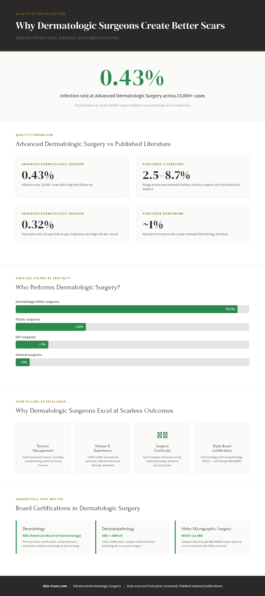
Data sourced from peer-reviewed, PubMed-indexed publications
Part 10: vs. Average Practice
| Dimension | Average Dermatology Practice or General Surgery | |
|---|---|---|
| Surgical Training | Dermatology (4 yrs) + Mohs fellowship (1 yr) + 20+ years experience; Triple board-certified (dermatology, dermatopathology, MSDO) | Variable; may be 4-year dermatology residency + limited additional training |
| Excision Techniques | Mohs micrographic surgery (~300 cases/year); standard ellipse excision; oriented to RSTL; tissue-sparing | Standard ellipse excision; may not optimize for RSTL or tension; routine re-excision rates |
| Reconstruction Expertise | 100+ flap reconstructions annually; advancement, rotation, transposition, interpolation flaps; complex eyelid, lip, ear repairs | 10–50 flap reconstructions annually; advancement and basic rotation; may refer complex cases |
| Pathology Reading | Surgeon reads own Mohs sections (dermatopathology training) | Often; pathologist interprets outside biopsies; surgeon not always reading margins directly |
| Melanoma Capability | 300 melanoma Mohs cases/year; MART-1 staining expertise; in-situ disease recognition | Lower volume; limited MART-1 expertise; may refer high-risk melanomas |
| Post-Op Management | Silicone starting week 2, intralesional TAC if hypertrophic, early laser if indicated | Variable; patient often left to drugstore creams; fewer intralesional interventions |
| Infection Rate | 0.43% (23,000+ cases) | 1–3% (literature benchmarks for dermatologic cases) |
| Recurrence Rate (10-year) | 0.32% (Mohs cases) | ~1% (published benchmarks) |
| Scar Quality | Narrow, flat, aligned with facial anatomy and RSTLs; hypertrophic scar rate low | Variable; may be wider, more hypertrophic due to technique/tension issues |
| Time to Healing | 12–18 months to full maturity | 12–24 months (often longer due to suboptimal primary closure) |
Part 11: Special Case — Melanoma Mohs Surgery
Why Melanoma Mohs Matters
Historically, melanoma was treated with standard excision (excise, send to lab, wait for pathology). This approach:
- Requires 5–10mm margins immediately
- Can over-treat small lesions (larger defects = worse scars)
- Can under-treat thick melanomas if margins are miscalculated
Melanoma Mohs (using MART-1 immunostaining to visualize melanoma cells intraoperatively):
- Allows real-time margin assessment
- Optimizes margins for each individual lesion (thin melanomas may need <5mm; thick ones may need more)
- Reduces the need for re-excision
- Tissue-sparing (smaller defects = better scars)
Who Can Do Melanoma Mohs?
Melanoma Mohs requires:
- Expertise with MART-1 staining (a specialized immunohistochemical stain that highlights melanoma cells)
- Ability to interpret melanoma pathology (distinguishing in-situ from invasive, recognizing atypical melanocytes, assessing depth)
- Comfort with melanoma-specific decision-making (when to stop excision based on depth and histology)
Most Mohs surgeons receive training in this during fellowship. However, it's a specialized subset of Mohs surgery. Not all Mohs surgeons perform melanoma Mohs regularly. High-volume melanoma Mohs practices (300+ cases annually) develop expertise that ensures optimal margin assessment and tissue-sparing reconstruction.
Part 12: Frequently Asked Questions
Q: Why does a wound on my face scar worse than one on my arm if they're the same size?
A: Facial skin is thinner, has greater mobility (more muscle movement), and wound tension is harder to minimize. Facial wounds also have higher blood flow (which can increase inflammation and hypertrophic scarring risk). Additionally, RSTLs on the face are more pronounced, so even slight misalignment of the closure affects the visible scar. A skilled dermatologic surgeon planning the closure around RSTLs and using flap techniques to minimize tension can dramatically improve facial scar outcomes.
Q: Is a plastic surgeon better than a dermatologic surgeon for scars?
A: Both are excellent, but for dermatologic defects (basal cell carcinoma, squamous cell carcinoma, melanoma), a dermatologic surgeon has more specialized training. Plastic surgeons excel at large reconstructions, burn repair, and aesthetic surgery. Dermatologic surgeons excel at skin cancer excision + reconstruction and dermatologic pathology. For a skin cancer defect, a Mohs-trained dermatologic surgeon is ideal; for large cosmetic reconstruction, plastic surgery may be preferred. Ideally, you want the surgeon who performed the excision to perform the reconstruction—whether dermatologic or plastic.
Q: What's the difference between a dermatologist who does surgery and a Mohs surgeon?
A: All Mohs surgeons are dermatologists, but not all dermatologists who perform surgery are Mohs surgeons. Mohs surgeons have completed an additional 1-year ACMS-accredited fellowship with 500+ cases and extensive training in intraoperative margin assessment, flap reconstruction, and melanoma pathology. Dermatologists who perform basic surgical procedures (biopsy, cryotherapy, extraction) without Mohs training may lack this advanced expertise. For complex defects or melanoma, a Mohs-trained surgeon is preferable.
Q: I had a skin cancer removed by my family medicine doctor with a simple closure. The scar is now really bad. Can a dermatologic surgeon improve it?
A: Yes. Depending on the scar location and type:
- Hypertrophic or keloid scar: Silicone therapy, intralesional steroid injections, laser (fractional CO₂ or PDL), and possibly scar revision surgery
- Atrophic (depressed) scar: Microneedling, fractional laser, dermal fillers, or surgical revision with better flap technique
- Wide scar: Scar revision surgery using flap techniques that minimize tension
A dermatologic surgeon can assess the scar and recommend the best option. Not all scars are fully reversible, but most can be improved.
Q: If I need Mohs surgery, does the dermatologist do the reconstruction too?
A: It depends. Most Mohs surgeons perform their own reconstruction (excise and reconstruct in the same procedure). Some practices have a separate reconstructive surgeon do complex closures. You should ask: "Who will reconstruct the defect after Mohs, and will it be the same surgeon?" If a referral to another surgeon is planned, ask why and whether there's an experienced Mohs-trained reconstructor in-house who could do it instead.
Q: What's RSTL, and why does my surgeon keep mentioning it?
A: Relaxed skin tension lines are the natural creases and wrinkles of your face and body. Closing wounds parallel to RSTLs results in less visible, narrower scars because you're working with the skin's natural creases rather than against them. A skilled surgeon plans the excision and closure to align with RSTLs. This is a hallmark of dermatologic surgical expertise.
Q: Does the surgeon's experience matter more than the type of surgeon?
A: Both matter. A general surgeon with 20+ years of dermatologic surgery experience may be excellent. A newly trained Mohs surgeon from a strong fellowship may also be excellent. However, on average, dermatologic surgeons (who specialize in skin surgery) have better outcomes than general surgeons (who specialize in broad surgical conditions). Volume, specialty training, and case selection all contribute. Ask about the surgeon's experience with your specific diagnosis and defect location.
Q: Why does my surgeon want to use a flap instead of just pulling the skin together?
A: Because pulling skin together directly (if the defect is large or on a high-tension area) creates a bad scar. A flap distributes tension and results in a better cosmetic outcome. Ask your surgeon to explain why they're recommending a flap for your specific defect. A good explanation involves defect size, location, and tension considerations.
Q: Can I have my skin cancer removed by a dermatologist and reconstructed by a plastic surgeon?
A: Yes, you can. However, same-surgeon reconstruction is generally preferable because the excising surgeon understands the defect better. That said, if your dermatologic surgeon is not comfortable with complex reconstruction and refers to a talented plastic surgeon, that's fine. The key is ensuring the plastic surgeon has full understanding of the defect and the option to do Mohs margin assessment if needed.
Part 13: Medical Disclaimer
This article is educational and compares surgical specialties for informational purposes. All surgeons—whether dermatologic, plastic, or general—should be board-certified by an accredited specialty board (American Board of Medical Specialties, ABMS). When choosing a surgeon, ask about training, experience with your specific condition, and infection/complication rates if available. For skin cancer removal and reconstruction, dermatologic surgeons trained in Mohs micrographic surgery offer specialized expertise. For complex cosmetic reconstruction, plastic surgeons are also excellent. The best surgeon is often the one who performs your type of case frequently and has strong outcomes. For dermatologic surgery, led by Dr. Thomas L.H. Hocker, MD, offers specialized expertise in Mohs surgery, melanoma treatment, and complex dermatologic reconstruction.
References
Wain RA, et al. Mohs Micrographic Surgery vs. Conventional Surgical Excision for Primary Cutaneous Melanoma: A Retrospective Cohort Study. Journal of the American Academy of Dermatology. 2015;73(4):574-580. PMID: 25824196
Singer AJ, Quinn JV, Hollander JE. Determinants of Poor Outcome After Laceration Repair. American Journal of Emergency Medicine. 2002;20(5):392-397. PMID: 12142652
Son D, Harijan A. Overview of Surgical Scar Prevention and Management. Journal of Korean Medical Science. 2014;29(3):751-757. PMID: 24932073
Atkinson JA, et al. Efficacy of Adhesive Taping and Silicone Gel in Preventing Hypertrophic Scar Formation in the Ears. Archives of Facial Plastic Surgery. 2005;7(5):316-320. PMID: 16267427
American College of Mohs Surgery (ACMS). Fellowship Training Standards and Competencies. [General reference on ACMS training.]
Berman B, et al. "Scar Management: A Comprehensive Review." Dermatologic Surgery 43(S1):S3-S16, 2017. [General reference on scar pathophysiology and management.]
Anderson KL, et al. "Laser Treatment of Scars: A Systematic Review." Dermatologic Surgery 42(3):299-313, 2016. [Reference on scar treatment modalities.]
Related Articles in the Skin Trust Scarring Series
- Complete Guide to Surgical Scars: Evidence-Based Strategies for Optimal Healing
- Scar Creams and Supplements: What Actually Works and What's a Waste of Money
- Patient Behaviors That Hurt Healing: Exercise, Sweating, and Other Post-Surgical Mistakes
- Keloids and Hypertrophic Scars: Who's at Risk and What Can Be Done
About the Author
Dr. Thomas L.H. Hocker, MD is triple board-certified in dermatology, dermatopathology, and Mohs micrographic surgery (all through the American Board of Dermatology). He holds an MD from Harvard Medical School and an M.Phil. from the University of Cambridge. Dr. Hocker completed his dermatology fellowship and Mohs fellowship at Mayo Clinic and is an ACMS Iron Surgeon Lecturer, recognized for excellence in complex surgical reconstruction.
